 Movie
Movie Controller

[English] 日本語
 Yorodumi
- PDB-1ob0: Kinetic stabilization of Bacillus licheniformis alpha-amylase thr... -
+ Open data

- Basic information
| Entry | Database: PDB / ID: 1ob0 | ||||||
|---|---|---|---|---|---|---|---|
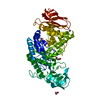
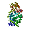
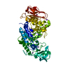
| Title | Kinetic stabilization of Bacillus licheniformis alpha-amylase through introduction of hydrophobic residues at the surface | ||||||
 Components Components | ALPHA-AMYLASE | ||||||
 Keywords Keywords | HYDROLASE / GLYCOSYLTRANSFERASE / STARCH DEGRADATION / THERMOSTABILITY / CALCIUM / SODIUM | ||||||
| Function / homology |  Function and homology informationalpha-amylase / alpha-amylase activity / carbohydrate metabolic process / calcium ion binding / extracellular space Function and homology informationalpha-amylase / alpha-amylase activity / carbohydrate metabolic process / calcium ion binding / extracellular spaceSimilarity search - Function | ||||||
| Biological species |  BACILLUS LICHENIFORMIS (bacteria) BACILLUS LICHENIFORMIS (bacteria) | ||||||
| Method | X-RAY DIFFRACTION / MOLECULAR REPLACEMENT / Resolution: 1.83 Å | ||||||
 Authors Authors | Machius, M. / Declerck, N. / Huber, R. / Wiegand, G. | ||||||
 Citation Citation | Journal: J.Biol.Chem. / Year: 2003 Title: Kinetic Stabilization of Bacillus Licheniformis Alpha-Amylase Through Introduction of Hydrophobic Residues at the Surface Authors: Machius, M. / Declerck, N. / Huber, R. / Wiegand, G. #1: Journal: Mol.Cells / Year: 1997Title: Crystal Structure of Thermostable Alpha-Amylase from Bacillus Licheniformis Refined at 1.7 A Resolution Authors: Hwang, K.Y. / Song, H.K. / Chang, C. / Lee, J. / Lee, S.Y. / Kim, K.K. / Choe, S. / Sweet, R.M. / Suh, S.W. #2: Journal: Structure / Year: 1998Title: Activation of Bacillus Licheniformis Alpha-Amylase Through a Disorder-->Order Transition of the Substrate-Binding Site Mediated by a Calcium-Sodium-Calcium Metal Triad Authors: Machius, M. / Declerck, N. / Huber, R. / Wiegand, G. #3: Journal: J.Mol.Biol. / Year: 1995Title: Crystal Structure of Calcium-Depleted Bacillus Licheniformis Alpha-Amylase at 2.2 A Resolution Authors: Machius, M. / Wiegand, G. / Huber, R. | ||||||
| History |
| ||||||
| Remark 700 | SHEET DETERMINATION METHOD: DSSP THE SHEETS PRESENTED AS "AA" IN EACH CHAIN ON SHEET RECORDS BELOW ... SHEET DETERMINATION METHOD: DSSP THE SHEETS PRESENTED AS "AA" IN EACH CHAIN ON SHEET RECORDS BELOW IS ACTUALLY AN 9-STRANDED BARREL THIS IS REPRESENTED BY A 10-STRANDED SHEET IN WHICH THE FIRST AND LAST STRANDS ARE IDENTICAL. SHEET THE SHEET STRUCTURE OF THIS MOLECULE IS BIFURCATED. IN ORDER TO REPRESENT THIS FEATURE IN THE SHEET RECORDS BELOW, TWO SHEETS ARE DEFINED. |
- Structure visualization
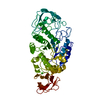
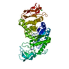
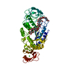
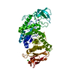
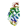
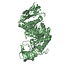
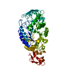
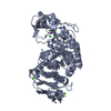
| Structure viewer | Molecule: MolmilJmol/JSmol |
|---|
- Downloads & links
-Download
| PDBx/mmCIF format | 1ob0.cif.gz | 123.5 KB | Display | PDBx/mmCIF format |
|---|---|---|---|---|
| PDB format | pdb1ob0.ent.gz | 95.1 KB | Display | PDB format |
| PDBx/mmJSON format | 1ob0.json.gz | Tree view | PDBx/mmJSON format | |
| Others |  Other downloads Other downloads |
-Validation report
| Arichive directory | https://data.pdbj.org/pub/pdb/validation_reports/ob/1ob0ftp://data.pdbj.org/pub/pdb/validation_reports/ob/1ob0 | HTTPS FTP |
|---|
-Related structure data
| Related structure data | 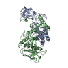 1bplS S: Starting model for refinement |
|---|---|
| Similar structure data |


-Links
 PDBj
PDBj

- Assembly
| Deposited unit | 
| ||||||||
|---|---|---|---|---|---|---|---|---|---|
| 1 |
| ||||||||
| Unit cell |
|
-Components
| #1: Protein | / 1 / 4-ALPHA-D-GLUCAN-4-GLUCANOHYDROLASE Mass: 55314.027 Da / Num. of mol.: 1 / Mutation: YES Source method: isolated from a genetically manipulated source Source: (gene. exp.) BACILLUS LICHENIFORMIS (bacteria) / Production host: BACILLUS SUBTILIS (bacteria) / References: UniProt: P06278, alpha-amylase | ||||||||
|---|---|---|---|---|---|---|---|---|---|
| #2: Chemical |   Mass: 40.078 Da / Num. of mol.: 3 / Source method: obtained synthetically / Formula: Ca Mass: 40.078 Da / Num. of mol.: 3 / Source method: obtained synthetically / Formula: Ca#3: Chemical | ChemComp-NA / |   Mass: 22.990 Da / Num. of mol.: 1 / Source method: obtained synthetically / Formula: Na Mass: 22.990 Da / Num. of mol.: 1 / Source method: obtained synthetically / Formula: Na#4: Water | ChemComp-HOH / | Water Mass: 18.015 Da / Num. of mol.: 324 / Source method: isolated from a natural source / Formula: H2O Mass: 18.015 Da / Num. of mol.: 324 / Source method: isolated from a natural source / Formula: H2OCompound details | CATALYSES ENDOHYDROLYSIS OF 1,4-ALPHA-GLUCOSIDIC LINKAGES IN OLIGOSACCHARIDES AND POLYSACCHARIDES. ...CATALYSES ENDOHYDROL | Sequence details | DATABASE ENTRY SWS P06278 CONTAINS A 29 RESIDUE PRECURSOR. NUMBERING IN THE PDB ENTRY STARTS WITH 1 ...DATABASE ENTRY SWS P06278 CONTAINS A 29 RESIDUE PRECURSOR. NUMBERING IN THE PDB ENTRY STARTS WITH 1 WHICH CORRESPOND | |
-Experimental details
-Experiment
| Experiment | Method: X-RAY DIFFRACTION / Number of used crystals: 1 |
|---|
- Sample preparation
| Crystal | Density Matthews: 3 Å3/Da / Density % sol: 59 % | ||||||||||||||||||||||||||||||||||||
|---|---|---|---|---|---|---|---|---|---|---|---|---|---|---|---|---|---|---|---|---|---|---|---|---|---|---|---|---|---|---|---|---|---|---|---|---|---|
| Crystal grow | Method: vapor diffusion / pH: 7 Details: PROTEIN WAS CRYSTALLIZED BY VAPOR DIFFUSION FROM DROPS CONTAINING 4 UL OF PROTEIN SOLUTION (10 MG/ML IN 50 MM TRIS/HCL, PH 8.0) PLUS 4 UL OF RESERVOIR SOLUTION (50 MM HEPES, 1 M AMMONIUM ...Details: PROTEIN WAS CRYSTALLIZED BY VAPOR DIFFUSION FROM DROPS CONTAINING 4 UL OF PROTEIN SOLUTION (10 MG/ML IN 50 MM TRIS/HCL, PH 8.0) PLUS 4 UL OF RESERVOIR SOLUTION (50 MM HEPES, 1 M AMMONIUM SULFATE, 1% (V/V) PEG 500, PH 7.0) EQUILIBRATED AGAINST 1 ML OF RESERVOIR SOLUTION. | ||||||||||||||||||||||||||||||||||||
| Crystal grow | *PLUS Temperature: 20 ℃ / pH: 8 / Method: vapor diffusion | ||||||||||||||||||||||||||||||||||||
| Components of the solutions | *PLUS
|
-Data collection
| Diffraction | Mean temperature: 278 K |
|---|---|
| Diffraction source | Source: ROTATING ANODE / Type: RIGAKU RU200 / Wavelength: 1.5418 |
| Detector | Type: MARRESEARCH / Detector: IMAGE PLATE / Date: May 15, 1997 / Details: BEAM FOCUSSING MIRROR SYSTEM (MSC, USA) |
| Radiation | Monochromator: NI FILTER / Protocol: SINGLE WAVELENGTH / Monochromatic (M) / Laue (L): M / Scattering type: x-ray |
| Radiation wavelength | Wavelength: 1.5418 Å / Relative weight: 1 |
| Reflection | Resolution: 1.83→25.9 Å / Num. obs: 55286 / % possible obs: 96.9 % / Observed criterion σ(I): 0 / Redundancy: 4 % / Biso Wilson estimate: 19.7 Å2 / Rmerge(I) obs: 0.067 / Net I/σ(I): 19 |
| Reflection shell | Resolution: 1.83→1.85 Å / Redundancy: 2.6 % / Rmerge(I) obs: 0.444 / Mean I/σ(I) obs: 2.1 / % possible all: 96.1 |
| Reflection | *PLUS Highest resolution: 1.83 Å / Num. obs: 61355 / % possible obs: 83.8 % / Redundancy: 3.8 % / Num. measured all: 231592 / Rmerge(I) obs: 0.073 |
| Reflection shell | *PLUS Highest resolution: 1.7 Å / Lowest resolution: 1.79 Å / % possible obs: 25 % / Rmerge(I) obs: 0.31 |
- Processing
| Software |
| ||||||||||||||||||||||||||||||||||||||||||||||||||||||||||||||||||||||||||||||||
|---|---|---|---|---|---|---|---|---|---|---|---|---|---|---|---|---|---|---|---|---|---|---|---|---|---|---|---|---|---|---|---|---|---|---|---|---|---|---|---|---|---|---|---|---|---|---|---|---|---|---|---|---|---|---|---|---|---|---|---|---|---|---|---|---|---|---|---|---|---|---|---|---|---|---|---|---|---|---|---|---|---|
| Refinement | Method to determine structure: MOLECULAR REPLACEMENT Starting model: PDB ENTRY 1BPL Resolution: 1.83→25.89 Å / Rfactor Rfree error: 0.004 / Data cutoff high absF: 987507.06 / Data cutoff low absF: 0 / Isotropic thermal model: RESTRAINED / Cross valid method: THROUGHOUT / σ(F): 0
| ||||||||||||||||||||||||||||||||||||||||||||||||||||||||||||||||||||||||||||||||
| Solvent computation | Solvent model: FLAT MODEL / Bsol: 48.3911 Å2 / ksol: 0.357256 e/Å3 | ||||||||||||||||||||||||||||||||||||||||||||||||||||||||||||||||||||||||||||||||
| Displacement parameters | Biso mean: 23.7 Å2
| ||||||||||||||||||||||||||||||||||||||||||||||||||||||||||||||||||||||||||||||||
| Refine analyze |
| ||||||||||||||||||||||||||||||||||||||||||||||||||||||||||||||||||||||||||||||||
| Refinement step | Cycle: LAST / Resolution: 1.83→25.89 Å
| ||||||||||||||||||||||||||||||||||||||||||||||||||||||||||||||||||||||||||||||||
| Refine LS restraints |
| ||||||||||||||||||||||||||||||||||||||||||||||||||||||||||||||||||||||||||||||||
| LS refinement shell | Resolution: 1.83→1.94 Å / Rfactor Rfree error: 0.015 / Total num. of bins used: 6
| ||||||||||||||||||||||||||||||||||||||||||||||||||||||||||||||||||||||||||||||||
| Xplor file |
| ||||||||||||||||||||||||||||||||||||||||||||||||||||||||||||||||||||||||||||||||
| Refinement | *PLUS Highest resolution: 1.83 Å / Lowest resolution: 25.89 Å / Num. reflection obs: 58628 / Num. reflection Rfree: 2345 / % reflection Rfree: 5 % / Rfactor Rfree: 0.174 / Rfactor Rwork: 0.156 | ||||||||||||||||||||||||||||||||||||||||||||||||||||||||||||||||||||||||||||||||
| Solvent computation | *PLUS | ||||||||||||||||||||||||||||||||||||||||||||||||||||||||||||||||||||||||||||||||
| Displacement parameters | *PLUS | ||||||||||||||||||||||||||||||||||||||||||||||||||||||||||||||||||||||||||||||||
| Refine LS restraints | *PLUS
| ||||||||||||||||||||||||||||||||||||||||||||||||||||||||||||||||||||||||||||||||
| LS refinement shell | *PLUS Highest resolution: 1.7 Å / Lowest resolution: 1.73 Å / Rfactor Rfree: 0.33 / Rfactor Rwork: 0.319 |
