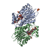
 Movie
Movie Controller


+ Open data

- Basic information
| Entry | Database: PDB / ID: 1oaa | ||||||
|---|---|---|---|---|---|---|---|
| Title | MOUSE SEPIAPTERIN REDUCTASE COMPLEXED WITH NADP AND OXALOACETATE | ||||||
 Components Components | SEPIAPTERIN REDUCTASE | ||||||
 Keywords Keywords | OXIDOREDUCTASE / SEPIAPTERIN REDUCTASE / TETRAHYDROBIOPTERIN | ||||||
| Function / homology |  Function and homology information Function and homology informationpteridine metabolic process / tetrahydrobiopterin metabolic process / sepiapterin reductase (L-erythro-7,8-dihydrobiopterin forming) / sepiapterin reductase activity / L-phenylalanine metabolic process / Tetrahydrobiopterin (BH4) synthesis, recycling, salvage and regulation / eNOS activation / cell morphogenesis involved in neuron differentiation / voluntary musculoskeletal movement / serotonin metabolic process ...pteridine metabolic process / tetrahydrobiopterin metabolic process / sepiapterin reductase (L-erythro-7,8-dihydrobiopterin forming) / sepiapterin reductase activity / L-phenylalanine metabolic process / Tetrahydrobiopterin (BH4) synthesis, recycling, salvage and regulation / eNOS activation / cell morphogenesis involved in neuron differentiation / voluntary musculoskeletal movement / serotonin metabolic process / tetrahydrobiopterin biosynthetic process / norepinephrine metabolic process / dopamine metabolic process / regulation of multicellular organism growth / protein homodimerization activity / mitochondrionSimilarity search - Function | ||||||
| Biological species |  Mus musculus (house mouse) Mus musculus (house mouse) | ||||||
| Method | X-RAY DIFFRACTION / SYNCHROTRON / MIR / Resolution: 1.25 Å | ||||||
 Authors Authors | Auerbach, G. / Herrmann, A. / Bacher, A. / Huber, R. | ||||||
 Citation Citation | Journal: EMBO J. / Year: 1997 Title: The 1.25 A crystal structure of sepiapterin reductase reveals its binding mode to pterins and brain neurotransmitters. Authors: Auerbach, G. / Herrmann, A. / Gutlich, M. / Fischer, M. / Jacob, U. / Bacher, A. / Huber, R. #1: Journal: Biol.Chem. / Year: 1997Title: The Pathway from GTP to Tetrahydrobiopterin: Three-Dimensional Structures of GTP Cyclohydrolase I and 6-Pyruvoyl Tetrahydropterin Synthase Authors: Auerbach, G. / Nar, H. #2: Journal: J.Mol.Biol. / Year: 1995Title: 6-Pyruvoyl Tetrahydropterin Synthase, an Enzyme with a Novel Type of Active Site Involving Both Zinc Binding and an Intersubunit Catalytic Triad Motif; Site-Directed Mutagenesis of the ...Title: 6-Pyruvoyl Tetrahydropterin Synthase, an Enzyme with a Novel Type of Active Site Involving Both Zinc Binding and an Intersubunit Catalytic Triad Motif; Site-Directed Mutagenesis of the Proposed Active Center, Characterization of the Metal Binding Site and Modelling of Substrate Binding Authors: Burgisser, D.M. / Thony, B. / Redweik, U. / Hess, D. / Heizmann, C.W. / Huber, R. / Nar, H. #3: Journal: Proc.Natl.Acad.Sci.USA / Year: 1995Title: Active Site Topology and Reaction Mechanism of GTP Cyclohydrolase I Authors: Nar, H. / Huber, R. / Auerbach, G. / Fischer, M. / Hosl, C. / Ritz, H. / Bracher, A. / Meining, W. / Eberhardt, S. / Bacher, A. #4: Journal: Structure / Year: 1995Title: Atomic Structure of GTP Cyclohydrolase I Authors: Nar, H. / Huber, R. / Meining, W. / Schmid, C. / Weinkauf, S. / Bacher, A. #5: Journal: Embo J. / Year: 1994Title: Three-Dimensional Structure of 6-Pyruvoyl Tetrahydropterin Synthase, an Enzyme Involved in Tetrahydrobiopterin Biosynthesis Authors: Nar, H. / Huber, R. / Heizmann, C.W. / Thony, B. / Burgisser, D. | ||||||
| History |
|
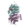
- Structure visualization
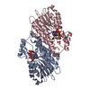
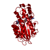
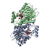
| Structure viewer | Molecule: MolmilJmol/JSmol |
|---|
- Downloads & links
-Download
| PDBx/mmCIF format | 1oaa.cif.gz | 77.1 KB | Display | PDBx/mmCIF format |
|---|---|---|---|---|
| PDB format | pdb1oaa.ent.gz | 55.9 KB | Display | PDB format |
| PDBx/mmJSON format | 1oaa.json.gz | Tree view | PDBx/mmJSON format | |
| Others |  Other downloads Other downloads |
-Validation report
| Arichive directory | https://data.pdbj.org/pub/pdb/validation_reports/oa/1oaaftp://data.pdbj.org/pub/pdb/validation_reports/oa/1oaa | HTTPS FTP |
|---|
-Related structure data


-Links
 PDBj
PDBj


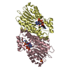
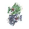
- Assembly
| Deposited unit | 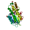
| ||||||||
|---|---|---|---|---|---|---|---|---|---|
| 1 | 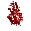
| ||||||||
| Unit cell |
| ||||||||
| Components on special symmetry positions |
|
-Components
| #1: Protein | Mass: 27648.684 Da / Num. of mol.: 1 Source method: isolated from a genetically manipulated source Source: (gene. exp.) Mus musculus (house mouse) / Organ: BRAIN / Cellular location (production host): CYTOSOL / Production host:  Escherichia coli (E. coli) Escherichia coli (E. coli)References: UniProt: Q64105, sepiapterin reductase (L-erythro-7,8-dihydrobiopterin forming) | ||||||
|---|---|---|---|---|---|---|---|
| #2: Chemical | Sulfate  Mass: 96.063 Da / Num. of mol.: 2 / Source method: obtained synthetically / Formula: SO4 Mass: 96.063 Da / Num. of mol.: 2 / Source method: obtained synthetically / Formula: SO4#3: Chemical | ChemComp-OAA / | Oxaloacetic acid  Mass: 131.064 Da / Num. of mol.: 1 / Source method: obtained synthetically / Formula: C4H3O5 Mass: 131.064 Da / Num. of mol.: 1 / Source method: obtained synthetically / Formula: C4H3O5#4: Chemical | ChemComp-NAP / | Nicotinamide adenine dinucleotide phosphate  Mass: 743.405 Da / Num. of mol.: 1 / Source method: obtained synthetically / Formula: C21H28N7O17P3 Mass: 743.405 Da / Num. of mol.: 1 / Source method: obtained synthetically / Formula: C21H28N7O17P3#5: Water | ChemComp-HOH / | Water Mass: 18.015 Da / Num. of mol.: 467 / Source method: isolated from a natural source / Formula: H2O Mass: 18.015 Da / Num. of mol.: 467 / Source method: isolated from a natural source / Formula: H2O |
-Experimental details
-Experiment
| Experiment | Method: X-RAY DIFFRACTION / Number of used crystals: 1 |
|---|
- Sample preparation
| Crystal | Density Matthews: 3.66 Å3/Da / Density % sol: 66 % | |||||||||||||||||||||||||||||||||||
|---|---|---|---|---|---|---|---|---|---|---|---|---|---|---|---|---|---|---|---|---|---|---|---|---|---|---|---|---|---|---|---|---|---|---|---|---|
| Crystal grow | *PLUS Temperature: 4 ℃ / pH: 6.5 / Method: vapor diffusion | |||||||||||||||||||||||||||||||||||
| Components of the solutions | *PLUS
|
-Data collection
| Diffraction | Mean temperature: 95 K |
|---|---|
| Diffraction source | Source: SYNCHROTRON / Site: MPG/DESY, HAMBURG  / Beamline: BW6 / Wavelength: 1 / Beamline: BW6 / Wavelength: 1 |
| Detector | Type: MARRESEARCH / Detector: IMAGE PLATE / Date: Sep 1, 1996 |
| Radiation | Monochromatic (M) / Laue (L): M / Scattering type: x-ray |
| Radiation wavelength | Wavelength: 1 Å / Relative weight: 1 |
| Reflection | Resolution: 1.25→8 Å / Num. obs: 112800 / % possible obs: 99.6 % / Observed criterion σ(I): 0 / Redundancy: 2.9 % / Rmerge(I) obs: 0.068 |
| Reflection shell | Resolution: 1.25→1.28 Å / Redundancy: 2.9 % / % possible all: 97.3 |
| Reflection | *PLUS Num. measured all: 630228 |
| Reflection shell | *PLUS % possible obs: 97.3 % |
- Processing
| Software |
| ||||||||||||||||||||||||||||||||||||||||||||||||||||||||||||
|---|---|---|---|---|---|---|---|---|---|---|---|---|---|---|---|---|---|---|---|---|---|---|---|---|---|---|---|---|---|---|---|---|---|---|---|---|---|---|---|---|---|---|---|---|---|---|---|---|---|---|---|---|---|---|---|---|---|---|---|---|---|
| Refinement | Method to determine structure: MIR / Resolution: 1.25→6 Å / Cross valid method: FREE-R / σ(F): 0
| ||||||||||||||||||||||||||||||||||||||||||||||||||||||||||||
| Refinement step | Cycle: LAST / Resolution: 1.25→6 Å
| ||||||||||||||||||||||||||||||||||||||||||||||||||||||||||||
| Refine LS restraints |
| ||||||||||||||||||||||||||||||||||||||||||||||||||||||||||||
| Software | *PLUS Name: X-PLOR / Version: 3.851 / Classification: refinement | ||||||||||||||||||||||||||||||||||||||||||||||||||||||||||||
| Refinement | *PLUS | ||||||||||||||||||||||||||||||||||||||||||||||||||||||||||||
| Solvent computation | *PLUS | ||||||||||||||||||||||||||||||||||||||||||||||||||||||||||||
| Displacement parameters | *PLUS Biso mean: 29 Å2 |
