 Movie
Movie Controller


+ Open data

- Basic information
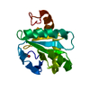
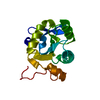
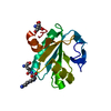
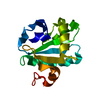
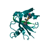
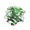
| Entry | Database: PDB / ID: 1o73 | ||||||
|---|---|---|---|---|---|---|---|
| Title | Tryparedoxin from Trypanosoma brucei | ||||||
 Components Components | TRYPAREDOXIN | ||||||
 Keywords Keywords |  ELECTRON TRANSPORT / TRYPAREDOXIN / TRYPANOSOMATID / TRYPANOSOMA BRUCEI / THIOREDOXIN ELECTRON TRANSPORT / TRYPAREDOXIN / TRYPANOSOMATID / TRYPANOSOMA BRUCEI / THIOREDOXIN | ||||||
| Function / homology |  Function and homology information Function and homology informationthioredoxin-disulfide reductase (NADPH) activity / negative regulation of Wnt signaling pathway / negative regulation of protein ubiquitination / nucleusSimilarity search - Function | ||||||
| Biological species |  TRYPANOSOMA BRUCEI BRUCEI (eukaryote) TRYPANOSOMA BRUCEI BRUCEI (eukaryote) | ||||||
| Method | X-RAY DIFFRACTION / MOLECULAR REPLACEMENT / Resolution: 2.28 Å | ||||||
 Authors Authors | Gabrielsen, M. / Alphey, M.S. / Bond, C.S. / Hunter, W.N. | ||||||
 Citation Citation | Journal: J.Biol.Chem. / Year: 2003 Title: Tryparedoxins from Crithidia Fasciculata and Trypanosoma Brucei: Photoreduction of the Redox Disulfide Using Synchrotron Radiation and Evidence for a Conformational Switch Implicated in Function Authors: Alphey, M.S. / Gabrielsen, M. / Micossi, E. / Leonard, G.A. / Mcsweeney, S.M. / Ravelli, R.B.G. / Tetaud, E. / Fairlamb, A.H. / Bond, C.S. / Hunter, W.N. | ||||||
| History |
|
- Structure visualization
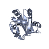
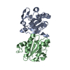
| Structure viewer | Molecule: MolmilJmol/JSmol |
|---|
- Downloads & links
-Download
| PDBx/mmCIF format | 1o73.cif.gz | 42.1 KB | Display | PDBx/mmCIF format |
|---|---|---|---|---|
| PDB format | pdb1o73.ent.gz | 28.6 KB | Display | PDB format |
| PDBx/mmJSON format | 1o73.json.gz | Tree view | PDBx/mmJSON format | |
| Others |  Other downloads Other downloads |
-Validation report
| Arichive directory | https://data.pdbj.org/pub/pdb/validation_reports/o7/1o73ftp://data.pdbj.org/pub/pdb/validation_reports/o7/1o73 | HTTPS FTP |
|---|
-Related structure data
| Related structure data |  1o7uC 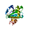 1o85C 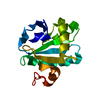 1o8wC  1o8xC  1oc8C  1oc9C 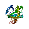 1qk8S C: citing same article ( S: Starting model for refinement |
|---|---|
| Similar structure data |


-Links
 PDBj
PDBj

- Assembly
| Deposited unit | 
| ||||||||
|---|---|---|---|---|---|---|---|---|---|
| 1 |
| ||||||||
| Unit cell |
|
-Components
| #1: Protein | Mass: 15904.936 Da / Num. of mol.: 1 Source method: isolated from a genetically manipulated source Source: (gene. exp.) TRYPANOSOMA BRUCEI BRUCEI (eukaryote) / Strain: 427 / Plasmid: PET-15B / Production host:  ESCHERICHIA COLI (E. coli) / Strain (production host): BL21 / References: UniProt: O77404 ESCHERICHIA COLI (E. coli) / Strain (production host): BL21 / References: UniProt: O77404 |
|---|---|
| #2: Water | ChemComp-HOH / Water Mass: 18.015 Da / Num. of mol.: 63 / Source method: isolated from a natural source / Formula: H2O Mass: 18.015 Da / Num. of mol.: 63 / Source method: isolated from a natural source / Formula: H2O |
| Compound details | THIS PROTEIN PARTICIPATES IN VARIOUS REDOX REACTIONS BY THE REVERSIBLE OXIDATION OF ITS ACTIVE ...THIS PROTEIN PARTICIPAT |
-Experimental details
-Experiment
| Experiment | Method: X-RAY DIFFRACTION / Number of used crystals: 1 |
|---|
- Sample preparation
| Crystal | Density Matthews: 2.5 Å3/Da / Density % sol: 50 % | ||||||||||||||||||||||||||||||||||||
|---|---|---|---|---|---|---|---|---|---|---|---|---|---|---|---|---|---|---|---|---|---|---|---|---|---|---|---|---|---|---|---|---|---|---|---|---|---|
| Crystal grow | pH: 4.6 Details: 50 MM HEPES PH 7.5, 30% PEG4000, 100 MM SODIUM ACETATE PH 4.6, 200 MM AMMONIUM ACETATE | ||||||||||||||||||||||||||||||||||||
| Crystal grow | *PLUS pH: 7.5 / Method: vapor diffusion, hanging drop | ||||||||||||||||||||||||||||||||||||
| Components of the solutions | *PLUS
|
-Data collection
| Diffraction | Mean temperature: 100 K |
|---|---|
| Diffraction source | Source: ROTATING ANODE / Type: RIGAKU RU200 / Wavelength: 1.5418 |
| Detector | Type: RIGAKU IMAGE PLATE / Detector: IMAGE PLATE / Details: ROTATING ANODE |
| Radiation | Protocol: SINGLE WAVELENGTH / Monochromatic (M) / Laue (L): M / Scattering type: x-ray |
| Radiation wavelength | Wavelength: 1.5418 Å / Relative weight: 1 |
| Reflection | Resolution: 2.28→56.8 Å / Num. obs: 5030 / % possible obs: 98.7 % / Observed criterion σ(I): 2 / Redundancy: 8.1 % / Rmerge(I) obs: 0.073 / Net I/σ(I): 19.6 |
| Reflection shell | Resolution: 2.28→2.36 Å / Redundancy: 3.4 % / Rmerge(I) obs: 0.217 / Mean I/σ(I) obs: 6.5 / % possible all: 94.7 |
| Reflection | *PLUS Highest resolution: 2.3 Å / Num. obs: 4699 / Num. measured all: 37965 |
| Reflection shell | *PLUS % possible obs: 94.7 % |
- Processing
| Software |
| ||||||||||||||||||||||||||||||||||||||||||||||||||||||||||||||||||||||||||||||||||||||||||||||||||||||||||||||||||||||||||||||||||||||||||||||||||||||||||||||||||||||||||||||||||||||
|---|---|---|---|---|---|---|---|---|---|---|---|---|---|---|---|---|---|---|---|---|---|---|---|---|---|---|---|---|---|---|---|---|---|---|---|---|---|---|---|---|---|---|---|---|---|---|---|---|---|---|---|---|---|---|---|---|---|---|---|---|---|---|---|---|---|---|---|---|---|---|---|---|---|---|---|---|---|---|---|---|---|---|---|---|---|---|---|---|---|---|---|---|---|---|---|---|---|---|---|---|---|---|---|---|---|---|---|---|---|---|---|---|---|---|---|---|---|---|---|---|---|---|---|---|---|---|---|---|---|---|---|---|---|---|---|---|---|---|---|---|---|---|---|---|---|---|---|---|---|---|---|---|---|---|---|---|---|---|---|---|---|---|---|---|---|---|---|---|---|---|---|---|---|---|---|---|---|---|---|---|---|---|---|
| Refinement | Method to determine structure: MOLECULAR REPLACEMENT Starting model: PDB ENTRY 1QK8 Resolution: 2.28→56.8 Å / Cor.coef. Fo:Fc: 0.944 / Cor.coef. Fo:Fc free: 0.902 / SU B: 8.827 / SU ML: 0.215 / Cross valid method: THROUGHOUT / ESU R: 0.802 / ESU R Free: 0.275 / Stereochemistry target values: MAXIMUM LIKELIHOOD / Details: HYDROGENS HAVE BEEN ADDED IN THE RIDING POSITIONS
| ||||||||||||||||||||||||||||||||||||||||||||||||||||||||||||||||||||||||||||||||||||||||||||||||||||||||||||||||||||||||||||||||||||||||||||||||||||||||||||||||||||||||||||||||||||||
| Solvent computation | Ion probe radii: 0.8 Å / Shrinkage radii: 0.8 Å / VDW probe radii: 1.4 Å / Solvent model: BABINET MODEL PLUS MASK | ||||||||||||||||||||||||||||||||||||||||||||||||||||||||||||||||||||||||||||||||||||||||||||||||||||||||||||||||||||||||||||||||||||||||||||||||||||||||||||||||||||||||||||||||||||||
| Displacement parameters | Biso mean: 26.62 Å2
| ||||||||||||||||||||||||||||||||||||||||||||||||||||||||||||||||||||||||||||||||||||||||||||||||||||||||||||||||||||||||||||||||||||||||||||||||||||||||||||||||||||||||||||||||||||||
| Refinement step | Cycle: LAST / Resolution: 2.28→56.8 Å
| ||||||||||||||||||||||||||||||||||||||||||||||||||||||||||||||||||||||||||||||||||||||||||||||||||||||||||||||||||||||||||||||||||||||||||||||||||||||||||||||||||||||||||||||||||||||
| Refine LS restraints |
|