 Movie
Movie Controller

[English] 日本語
 Yorodumi
- PDB-1o5w: The structure basis of specific recognitions for substrates and i... -
+ Open data

- Basic information
| Entry | Database: PDB / ID: 1o5w | ||||||
|---|---|---|---|---|---|---|---|
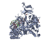
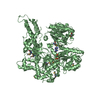
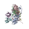
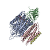
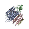
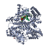
| Title | The structure basis of specific recognitions for substrates and inhibitors of rat monoamine oxidase A | ||||||
 Components Components | Amine oxidase [flavin-containing] A | ||||||
 Keywords Keywords |  OXIDOREDUCTASE OXIDOREDUCTASE | ||||||
| Function / homology |  Function and homology information Function and homology informationphenylethylamine metabolic process / Biogenic amines are oxidatively deaminated to aldehydes by MAOA and MAOB / : / Metabolism of serotonin / monoamine oxidase / positive regulation of signal transduction / Norepinephrine Neurotransmitter Release Cycle / Enzymatic degradation of dopamine by COMT / Enzymatic degradation of Dopamine by monoamine oxidase / catecholamine catabolic process ...phenylethylamine metabolic process / Biogenic amines are oxidatively deaminated to aldehydes by MAOA and MAOB / : / Metabolism of serotonin / monoamine oxidase / positive regulation of signal transduction / Norepinephrine Neurotransmitter Release Cycle / Enzymatic degradation of dopamine by COMT / Enzymatic degradation of Dopamine by monoamine oxidase / catecholamine catabolic process / serotonin metabolic process / dopamine catabolic process / serotonin binding / primary amine oxidase activity / flavin adenine dinucleotide binding / mitochondrial outer membrane / membrane => GO:0016020Similarity search - Function | ||||||
| Biological species |  Rattus norvegicus (Norway rat) Rattus norvegicus (Norway rat) | ||||||
| Method | X-RAY DIFFRACTION / MOLECULAR REPLACEMENT / Resolution: 3.2 Å | ||||||
 Authors Authors | Ma, J. / Yoshimura, M. / Yamashita, E. / Nakagawa, A. / Ito, A. / Tsukihara, T. | ||||||
 Citation Citation | Journal: J.Mol.Biol. / Year: 2004 Title: Structure of rat monoamine oxidase a and its specific recognitions for substrates and inhibitors. Authors: Ma, J. / Yoshimura, M. / Yamashita, E. / Nakagawa, A. / Ito, A. / Tsukihara, T. | ||||||
| History |
|
- Structure visualization
| Structure viewer | Molecule: MolmilJmol/JSmol |
|---|
- Downloads & links
-Download
| PDBx/mmCIF format | 1o5w.cif.gz | 422.1 KB | Display | PDBx/mmCIF format |
|---|---|---|---|---|
| PDB format | pdb1o5w.ent.gz | 341.3 KB | Display | PDB format |
| PDBx/mmJSON format | 1o5w.json.gz | Tree view | PDBx/mmJSON format | |
| Others |  Other downloads Other downloads |
-Validation report
| Arichive directory | https://data.pdbj.org/pub/pdb/validation_reports/o5/1o5wftp://data.pdbj.org/pub/pdb/validation_reports/o5/1o5w | HTTPS FTP |
|---|
-Related structure data
| Similar structure data |
|---|

-Links
 PDBj
PDBj

- Assembly
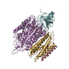
| Deposited unit | 
| ||||||||
|---|---|---|---|---|---|---|---|---|---|
| 1 | 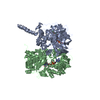
| ||||||||
| 2 | 
| ||||||||
| Unit cell |
|
-Components
| #1: Protein | Mass: 60613.883 Da / Num. of mol.: 4 Source method: isolated from a genetically manipulated source Source: (gene. exp.) Rattus norvegicus (Norway rat) / Plasmid: YEp51 / Production host:  Saccharomyces cerevisiae (brewer's yeast) / Strain (production host): BJ2168 / References: UniProt: P21396, monoamine oxidase Saccharomyces cerevisiae (brewer's yeast) / Strain (production host): BJ2168 / References: UniProt: P21396, monoamine oxidase#2: Chemical | ChemComp-FAD / Flavin adenine dinucleotide  Mass: 785.550 Da / Num. of mol.: 4 / Source method: obtained synthetically / Formula: C27H33N9O15P2 / Comment: FAD*YM Mass: 785.550 Da / Num. of mol.: 4 / Source method: obtained synthetically / Formula: C27H33N9O15P2 / Comment: FAD*YM#3: Chemical | ChemComp-MLG / Clorgiline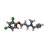  Mass: 272.170 Da / Num. of mol.: 4 / Source method: obtained synthetically / Formula: C13H15Cl2NO / Comment: antidepressant, inhibitor*YM Mass: 272.170 Da / Num. of mol.: 4 / Source method: obtained synthetically / Formula: C13H15Cl2NO / Comment: antidepressant, inhibitor*YM |
|---|
-Experimental details
-Experiment
| Experiment | Method: X-RAY DIFFRACTION |
|---|
- Sample preparation
| Crystal | Density Matthews: 3.47 Å3/Da / Density % sol: 64.31 % |
|---|---|
| Crystal grow | Method: vapor diffusion, hanging drop / Details: VAPOR DIFFUSION, HANGING DROP |
-Data collection
| Diffraction | Mean temperature: 100 K |
|---|---|
| Detector | Type: MACSCIENCE / Detector: IMAGE PLATE |
| Radiation | Protocol: SINGLE WAVELENGTH / Monochromatic (M) / Laue (L): M / Scattering type: x-ray |
| Radiation wavelength | Relative weight: 1 |
| Reflection | Num. obs: 59281 |
- Processing
| Software |
| ||||||||||||||||||||||||||||||||||||||||||||||||||||||||||||
|---|---|---|---|---|---|---|---|---|---|---|---|---|---|---|---|---|---|---|---|---|---|---|---|---|---|---|---|---|---|---|---|---|---|---|---|---|---|---|---|---|---|---|---|---|---|---|---|---|---|---|---|---|---|---|---|---|---|---|---|---|---|
| Refinement | Method to determine structure: MOLECULAR REPLACEMENT / Resolution: 3.2→16.19 Å / Rfactor Rfree error: 0.006 / Data cutoff high absF: 2848495.48 / Data cutoff low absF: 0 / Isotropic thermal model: RESTRAINED / Cross valid method: THROUGHOUT / σ(F): 0
| ||||||||||||||||||||||||||||||||||||||||||||||||||||||||||||
| Solvent computation | Solvent model: FLAT MODEL / Bsol: 42.7025 Å2 / ksol: 0.240749 e/Å3 | ||||||||||||||||||||||||||||||||||||||||||||||||||||||||||||
| Displacement parameters | Biso mean: 93.8 Å2
| ||||||||||||||||||||||||||||||||||||||||||||||||||||||||||||
| Refine analyze |
| ||||||||||||||||||||||||||||||||||||||||||||||||||||||||||||
| Refinement step | Cycle: LAST / Resolution: 3.2→16.19 Å
| ||||||||||||||||||||||||||||||||||||||||||||||||||||||||||||
| Refine LS restraints |
| ||||||||||||||||||||||||||||||||||||||||||||||||||||||||||||
| LS refinement shell | Resolution: 3.2→3.4 Å / Rfactor Rfree error: 0.021 / Total num. of bins used: 6
| ||||||||||||||||||||||||||||||||||||||||||||||||||||||||||||
| Xplor file |
|
