 Movie
Movie Controller

[English] 日本語
 Yorodumi
- PDB-1o5o: Crystal structure of Uracil phosphoribosyltransferase (TM0721) fr... -
+ Open data

- Basic information
| Entry | Database: PDB / ID: 1o5o | ||||||
|---|---|---|---|---|---|---|---|
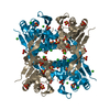
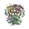
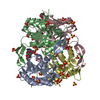
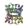
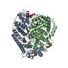
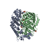
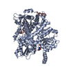
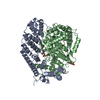
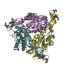
| Title | Crystal structure of Uracil phosphoribosyltransferase (TM0721) from Thermotoga maritima at 2.30 A resolution | ||||||
 Components Components | Uracil phosphoribosyltransferase | ||||||
 Keywords Keywords | TRANSFERASE / TM0721 / URACIL PHOSPHORIBOSYLTRANSFERASE / STRUCTURAL GENOMICS / JCSG / PSI / Protein Structure Initiative / Joint Center for Structural Genomics | ||||||
| Function / homology |  Function and homology information Function and homology informationuracil salvage / uracil phosphoribosyltransferase / uracil phosphoribosyltransferase activity / UMP salvage / nucleoside metabolic process / kinase activity / GTP binding / magnesium ion binding / cytosolSimilarity search - Function | ||||||
| Biological species |   Thermotoga maritima (bacteria) Thermotoga maritima (bacteria) | ||||||
| Method | X-RAY DIFFRACTION / SYNCHROTRON / MOLECULAR REPLACEMENT / Resolution: 2.3 Å | ||||||
 Authors Authors | Joint Center for Structural Genomics (JCSG) | ||||||
 Citation Citation | Journal: To be published Title: Crystal structure of Uracil phosphoribosyltransferase (TM0721) from Thermotoga maritima at 2.30 A resolution Authors: Joint Center for Structural Genomics (JCSG) | ||||||
| History |
|
- Structure visualization
| Structure viewer | Molecule: MolmilJmol/JSmol |
|---|
- Downloads & links
-Download
| PDBx/mmCIF format | 1o5o.cif.gz | 185.7 KB | Display | PDBx/mmCIF format |
|---|---|---|---|---|
| PDB format | pdb1o5o.ent.gz | 147.4 KB | Display | PDB format |
| PDBx/mmJSON format | 1o5o.json.gz | Tree view | PDBx/mmJSON format | |
| Others |  Other downloads Other downloads |
-Validation report
| Arichive directory | https://data.pdbj.org/pub/pdb/validation_reports/o5/1o5oftp://data.pdbj.org/pub/pdb/validation_reports/o5/1o5o | HTTPS FTP |
|---|
-Related structure data
| Related structure data |  1i5eS S: Starting model for refinement |
|---|---|
| Similar structure data | |
| Other databases |

-Links
 PDBj
PDBj


- Assembly
| Deposited unit | 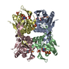
| |||||||||||||||||||||||||||||||||||||||||||||||||||||||||||||||||||||||||||||||||||||||||||||||||||||||||||||||||||||||||||||||||||||||||||||||||||||||||||||||||||||||||||||||||||||||||||||||||||||||||||||||||||||||||
|---|---|---|---|---|---|---|---|---|---|---|---|---|---|---|---|---|---|---|---|---|---|---|---|---|---|---|---|---|---|---|---|---|---|---|---|---|---|---|---|---|---|---|---|---|---|---|---|---|---|---|---|---|---|---|---|---|---|---|---|---|---|---|---|---|---|---|---|---|---|---|---|---|---|---|---|---|---|---|---|---|---|---|---|---|---|---|---|---|---|---|---|---|---|---|---|---|---|---|---|---|---|---|---|---|---|---|---|---|---|---|---|---|---|---|---|---|---|---|---|---|---|---|---|---|---|---|---|---|---|---|---|---|---|---|---|---|---|---|---|---|---|---|---|---|---|---|---|---|---|---|---|---|---|---|---|---|---|---|---|---|---|---|---|---|---|---|---|---|---|---|---|---|---|---|---|---|---|---|---|---|---|---|---|---|---|---|---|---|---|---|---|---|---|---|---|---|---|---|---|---|---|---|---|---|---|---|---|---|---|---|---|---|---|---|---|---|---|---|
| 1 |
| |||||||||||||||||||||||||||||||||||||||||||||||||||||||||||||||||||||||||||||||||||||||||||||||||||||||||||||||||||||||||||||||||||||||||||||||||||||||||||||||||||||||||||||||||||||||||||||||||||||||||||||||||||||||||
| Unit cell |
| |||||||||||||||||||||||||||||||||||||||||||||||||||||||||||||||||||||||||||||||||||||||||||||||||||||||||||||||||||||||||||||||||||||||||||||||||||||||||||||||||||||||||||||||||||||||||||||||||||||||||||||||||||||||||
| Noncrystallographic symmetry (NCS) | NCS domain:
NCS domain segments:
|