 Movie
Movie Controller

[English] 日本語
 Yorodumi
- PDB-1o58: Crystal structure of O-acetylserine sulfhydrylase (TM0665) from T... -
+ Open data

- Basic information
| Entry | Database: PDB / ID: 1o58 | |||||||||
|---|---|---|---|---|---|---|---|---|---|---|
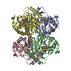
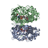
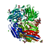
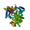
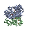
| Title | Crystal structure of O-acetylserine sulfhydrylase (TM0665) from Thermotoga maritima at 1.80 A resolution | |||||||||
 Components Components | O-acetylserine sulfhydrylase | |||||||||
 Keywords Keywords |  TRANSFERASE / TM0665 / O-ACETYLSERINE SULFHYDRYLASE / STRUCTURAL GENOMICS / JCSG / PSI / PROTEIN STRUCTURE INITIATIVE / JOINT CENTER FOR STRUCTURAL GENOMICS TRANSFERASE / TM0665 / O-ACETYLSERINE SULFHYDRYLASE / STRUCTURAL GENOMICS / JCSG / PSI / PROTEIN STRUCTURE INITIATIVE / JOINT CENTER FOR STRUCTURAL GENOMICS | |||||||||
| Function / homology |  Function and homology informationcysteine synthase / cysteine synthase activity / L-cysteine desulfhydrase activity / cysteine biosynthetic process from serine / pyridoxal phosphate binding / cytoplasm Function and homology informationcysteine synthase / cysteine synthase activity / L-cysteine desulfhydrase activity / cysteine biosynthetic process from serine / pyridoxal phosphate binding / cytoplasmSimilarity search - Function | |||||||||
| Biological species |   Thermotoga maritima (bacteria) Thermotoga maritima (bacteria) | |||||||||
| Method | X-RAY DIFFRACTION / SYNCHROTRON / MAD / Resolution: 1.8 Å | |||||||||
 Authors Authors | Joint Center for Structural Genomics (JCSG) | |||||||||
 Citation Citation | Journal: Proteins / Year: 2004 Title: Crystal structure of O-acetylserine sulfhydrylase (TM0665) from Thermotoga maritima at 1.8 A resolution Authors: Heine, A. / Canaves, J.M. / von Delft, F. / Brinen, L.S. / Dai, X. / Deacon, A.M. / Elsliger, M.A. / Eshaghi, S. / Floyd, R. / Godzik, A. / Grittini, C. / Grzechnik, S.K. / Guda, C. / ...Authors: Heine, A. / Canaves, J.M. / von Delft, F. / Brinen, L.S. / Dai, X. / Deacon, A.M. / Elsliger, M.A. / Eshaghi, S. / Floyd, R. / Godzik, A. / Grittini, C. / Grzechnik, S.K. / Guda, C. / Jaroszewski, L. / Karlak, C. / Klock, H.E. / Koesema, E. / Kovarik, J.S. / Kreusch, A. / Kuhn, P. / Lesley, S.A. / McMullan, D. / McPhillips, T.M. / Miller, M.A. / Miller, M.D. / Morse, A. / Moy, K. / Ouyang, J. / Page, R. / Robb, A. / Rodrigues, K. / Schwarzenbacher, R. / Selby, T.L. / Spraggon, G. / Stevens, R.C. / van den Bedem, H. / Velasquez, J. / Vincent, J. / Wang, X. / West, B. / Wolf, G. / Hodgson, K.O. / Wooley, J. / Wilson, I.A. | |||||||||
| History |
|
- Structure visualization
| Structure viewer | Molecule: MolmilJmol/JSmol |
|---|
- Downloads & links
-Download
| PDBx/mmCIF format | 1o58.cif.gz | 239.8 KB | Display | PDBx/mmCIF format |
|---|---|---|---|---|
| PDB format | pdb1o58.ent.gz | 192.4 KB | Display | PDB format |
| PDBx/mmJSON format | 1o58.json.gz | Tree view | PDBx/mmJSON format | |
| Others |  Other downloads Other downloads |
-Validation report
| Arichive directory | https://data.pdbj.org/pub/pdb/validation_reports/o5/1o58ftp://data.pdbj.org/pub/pdb/validation_reports/o5/1o58 | HTTPS FTP |
|---|
-Related structure data
| Similar structure data | |
|---|---|
| Other databases |


-Links
 PDBj
PDBj
- Assembly
| Deposited unit | 
| ||||||||||||||||||||||||||||||||||||||||||||||||||
|---|---|---|---|---|---|---|---|---|---|---|---|---|---|---|---|---|---|---|---|---|---|---|---|---|---|---|---|---|---|---|---|---|---|---|---|---|---|---|---|---|---|---|---|---|---|---|---|---|---|---|---|
| 1 |
| ||||||||||||||||||||||||||||||||||||||||||||||||||
| Unit cell |
| ||||||||||||||||||||||||||||||||||||||||||||||||||
| Noncrystallographic symmetry (NCS) | NCS domain:
NCS domain segments: Component-ID: 1 / Ens-ID: 1 / Beg auth comp-ID: HIS / Beg label comp-ID: HIS / Refine code: 6
|
-Components
| #1: Protein | Mass: 32631.938 Da / Num. of mol.: 4 Source method: isolated from a genetically manipulated source Source: (gene. exp.) Thermotoga maritima (bacteria) / Gene: TM0665 / Production host: Escherichia coli (E. coli) / References: UniProt: Q9WZD3, cysteine synthase#2: Chemical | ChemComp-PO4 / Phosphate  Mass: 94.971 Da / Num. of mol.: 8 / Source method: obtained synthetically / Formula: PO4 Mass: 94.971 Da / Num. of mol.: 8 / Source method: obtained synthetically / Formula: PO4#3: Water | ChemComp-HOH / | Water Mass: 18.015 Da / Num. of mol.: 937 / Source method: isolated from a natural source / Formula: H2O Mass: 18.015 Da / Num. of mol.: 937 / Source method: isolated from a natural source / Formula: H2O |
|---|
-Experimental details
-Experiment
| Experiment | Method: X-RAY DIFFRACTION / Number of used crystals: 1 |
|---|
- Sample preparation
| Crystal | Density Matthews: 2.32 Å3/Da / Density % sol: 46.5 % | |||||||||||||||||||||||||||||||||||
|---|---|---|---|---|---|---|---|---|---|---|---|---|---|---|---|---|---|---|---|---|---|---|---|---|---|---|---|---|---|---|---|---|---|---|---|---|
| Crystal grow | Temperature: 293 K / Method: vapor diffusion, sitting drop, nanodrop / pH: 9.2 Details: 50% PEG-400, 0.1M CHES pH 9.5, 0.2M NaCl, pH 9.2, VAPOR DIFFUSION,SITTING DROP,NANODROP, temperature 293K | |||||||||||||||||||||||||||||||||||
| Crystal grow | *PLUS pH: 7.9 / Method: vapor diffusion | |||||||||||||||||||||||||||||||||||
| Components of the solutions | *PLUS
|
-Data collection
| Diffraction | Mean temperature: 100 K | ||||||||||||
|---|---|---|---|---|---|---|---|---|---|---|---|---|---|
| Diffraction source | Source: SYNCHROTRON / Site: SSRL  / Beamline: BL9-2 / Wavelength: 0.97999, 0.97980, 0.91837 / Beamline: BL9-2 / Wavelength: 0.97999, 0.97980, 0.91837 | ||||||||||||
| Detector | Type: ADSC QUANTUM 315 / Detector: CCD / Date: Dec 10, 2001 | ||||||||||||
| Radiation | Monochromator: double crystal monochromator / Protocol: MAD / Monochromatic (M) / Laue (L): M / Scattering type: x-ray | ||||||||||||
| Radiation wavelength |
| ||||||||||||
| Reflection | Resolution: 1.8→48.66 Å / Num. all: 97122 / Num. obs: 97122 / % possible obs: 87.6 % / Redundancy: 4.1 % / Biso Wilson estimate: 29.14 Å2 / Rsym value: 0.038 / Net I/σ(I): 20.2 | ||||||||||||
| Reflection shell | Resolution: 1.8→1.85 Å / Redundancy: 3.2 % / Mean I/σ(I) obs: 4.3 / Num. unique all: 4897 / Rsym value: 0.273 / % possible all: 59.7 | ||||||||||||
| Reflection | *PLUS Highest resolution: 1.8 Å / Lowest resolution: 48.8 Å / Num. measured all: 400214 / Rmerge(I) obs: 0.038 | ||||||||||||
| Reflection shell | *PLUS % possible obs: 69.7 % / Rmerge(I) obs: 0.273 / Mean I/σ(I) obs: 2.6 |
- Processing
| Software |
| |||||||||||||||||||||||||||||||||||||||||||||||||||||||||||||||||||||||||||||||||||||||||||||||||||||||||||||||||||
|---|---|---|---|---|---|---|---|---|---|---|---|---|---|---|---|---|---|---|---|---|---|---|---|---|---|---|---|---|---|---|---|---|---|---|---|---|---|---|---|---|---|---|---|---|---|---|---|---|---|---|---|---|---|---|---|---|---|---|---|---|---|---|---|---|---|---|---|---|---|---|---|---|---|---|---|---|---|---|---|---|---|---|---|---|---|---|---|---|---|---|---|---|---|---|---|---|---|---|---|---|---|---|---|---|---|---|---|---|---|---|---|---|---|---|---|---|
| Refinement | Method to determine structure: MAD / Resolution: 1.8→48.66 Å / Cor.coef. Fo:Fc: 0.964 / Cor.coef. Fo:Fc free: 0.946 / SU B: 4.252 / SU ML: 0.069 / TLS residual ADP flag: LIKELY RESIDUAL / Isotropic thermal model: ISOTROPIC / Cross valid method: THROUGHOUT / ESU R: 0.125 / ESU R Free: 0.121 / Stereochemistry target values: MAXIMUM LIKELIHOOD Details: 1. THE STRAND-TURN-HELIX OF RESIDUES 102-126 IS FULLY ORDERED ONLY IN CHAIN A; WHAT IS INTERPRETABLE (VARIABLY) IN THE OTHER CHAINS DOES NOT OBEY NCS. IN PARTICULAR, STRONG DENSITY IS ...Details: 1. THE STRAND-TURN-HELIX OF RESIDUES 102-126 IS FULLY ORDERED ONLY IN CHAIN A; WHAT IS INTERPRETABLE (VARIABLY) IN THE OTHER CHAINS DOES NOT OBEY NCS. IN PARTICULAR, STRONG DENSITY IS VISIBLE AROUND THE ESTIMATED (BY NCS) RESIDUES C112-126 AND D109-118, BUT IS TOO AMBIGUOUS FOR INTERPRETATION. 2. DIFFERENCE DENSITY AROUND 83-85 IN CHAINS BCD INDICATE DISORDER, PRESUMABLY RELATED TO THE DISORDER OF ADJACENT RESIDUES 102-126 (POINT 1). 3. RESIDUES C60-61 DISOBEY NCS, BUT DIFFERENCE DENSITY INDICATES SUPERIMPOSED PRESENCE OF CONFORMATION FROM CHAINS ABD. LIKEWISE, DIFFERENCE DENSITY AT D60-61 INDICATES PRESENCE OF C CONFORMATION. NEITHER DENSITY WAS CLEAR ENOUGH TO BE MODELED AS ALTERNATIVE CONFORMATIONS. 4. HYDROGENS HAVE BEEN ADDED IN THE RIDING POSITIONS.
| |||||||||||||||||||||||||||||||||||||||||||||||||||||||||||||||||||||||||||||||||||||||||||||||||||||||||||||||||||
| Solvent computation | Ion probe radii: 0.8 Å / Shrinkage radii: 0.8 Å / VDW probe radii: 1.2 Å / Solvent model: BABINET MODEL WITH MASK | |||||||||||||||||||||||||||||||||||||||||||||||||||||||||||||||||||||||||||||||||||||||||||||||||||||||||||||||||||
| Displacement parameters | Biso mean: 22.156 Å2
| |||||||||||||||||||||||||||||||||||||||||||||||||||||||||||||||||||||||||||||||||||||||||||||||||||||||||||||||||||
| Refinement step | Cycle: LAST / Resolution: 1.8→48.66 Å
| |||||||||||||||||||||||||||||||||||||||||||||||||||||||||||||||||||||||||||||||||||||||||||||||||||||||||||||||||||
| Refine LS restraints |
| |||||||||||||||||||||||||||||||||||||||||||||||||||||||||||||||||||||||||||||||||||||||||||||||||||||||||||||||||||
| Refine LS restraints NCS | Ens-ID: 1 / Number: 3827 / Refine-ID: X-RAY DIFFRACTION
| |||||||||||||||||||||||||||||||||||||||||||||||||||||||||||||||||||||||||||||||||||||||||||||||||||||||||||||||||||
| LS refinement shell | Resolution: 1.8→1.847 Å / Total num. of bins used: 20
| |||||||||||||||||||||||||||||||||||||||||||||||||||||||||||||||||||||||||||||||||||||||||||||||||||||||||||||||||||
| Refinement TLS params. | Method: refined / Origin x: 41.501 Å / Origin y: 5.778 Å / Origin z: 80.26 Å
| |||||||||||||||||||||||||||||||||||||||||||||||||||||||||||||||||||||||||||||||||||||||||||||||||||||||||||||||||||
| Refinement TLS group |
| |||||||||||||||||||||||||||||||||||||||||||||||||||||||||||||||||||||||||||||||||||||||||||||||||||||||||||||||||||
| Refinement | *PLUS Num. reflection obs: 97093 / % reflection Rfree: 4.9 % / Rfactor Rfree: 0.198 / Rfactor Rwork: 0.157 | |||||||||||||||||||||||||||||||||||||||||||||||||||||||||||||||||||||||||||||||||||||||||||||||||||||||||||||||||||
| Solvent computation | *PLUS | |||||||||||||||||||||||||||||||||||||||||||||||||||||||||||||||||||||||||||||||||||||||||||||||||||||||||||||||||||
| Displacement parameters | *PLUS | |||||||||||||||||||||||||||||||||||||||||||||||||||||||||||||||||||||||||||||||||||||||||||||||||||||||||||||||||||
| Refine LS restraints | *PLUS
|
