 Movie
Movie Controller

[English] 日本語
 Yorodumi
- PDB-1o4v: Crystal structure of the catalytic subunit of a phosphoribosylami... -
+ Open data

- Basic information
| Entry | Database: PDB / ID: 1o4v | ||||||
|---|---|---|---|---|---|---|---|
| Title | Crystal structure of the catalytic subunit of a phosphoribosylaminoimidazole mutase (tm0446) from thermotoga maritima at 1.77 A resolution | ||||||
 Components Components | phosphoribosylaminoimidazole mutase PurE | ||||||
 Keywords Keywords |  LYASE / Structural genomics / Joint Center for Structural Genomics / JCSG / Protein Structure Initiative / PSI LYASE / Structural genomics / Joint Center for Structural Genomics / JCSG / Protein Structure Initiative / PSI | ||||||
| Function / homology |  Function and homology information5-(carboxyamino)imidazole ribonucleotide mutase / 5-(carboxyamino)imidazole ribonucleotide mutase activity / 'de novo' IMP biosynthetic process Function and homology information5-(carboxyamino)imidazole ribonucleotide mutase / 5-(carboxyamino)imidazole ribonucleotide mutase activity / 'de novo' IMP biosynthetic processSimilarity search - Function | ||||||
| Biological species |   Thermotoga maritima (bacteria) Thermotoga maritima (bacteria) | ||||||
| Method | X-RAY DIFFRACTION / SYNCHROTRON / MOLECULAR REPLACEMENT / Resolution: 1.77 Å | ||||||
 Authors Authors | Joint Center for Structural Genomics (JCSG) | ||||||
 Citation Citation | Journal: Proteins / Year: 2004 Title: Crystal structure of a phosphoribosylaminoimidazole mutase PurE (TM0446) from Thermotoga maritima at 1.77 A resolution Authors: Schwarzenbacher, R. / Jaroszewski, L. / von Delft, F. / Abdubek, P. / Ambing, E. / Biorac, T. / Brinen, L.S. / Canaves, J.M. / Cambell, J. / Chiu, H.J. / Dai, X. / Deacon, A.M. / DiDonato, M. ...Authors: Schwarzenbacher, R. / Jaroszewski, L. / von Delft, F. / Abdubek, P. / Ambing, E. / Biorac, T. / Brinen, L.S. / Canaves, J.M. / Cambell, J. / Chiu, H.J. / Dai, X. / Deacon, A.M. / DiDonato, M. / Elsliger, M.A. / Eshagi, S. / Floyd, R. / Godzik, A. / Grittini, C. / Grzechnik, S.K. / Hampton, E. / Karlak, C. / Klock, H.E. / Koesema, E. / Kovarik, J.S. / Kreusch, A. / Kuhn, P. / Lesley, S.A. / Levin, I. / McMullan, D. / McPhillips, T.M. / Miller, M.D. / Morse, A. / Moy, K. / Ouyang, J. / Page, R. / Quijano, K. / Robb, A. / Spraggon, G. / Stevens, R.C. / van den Bedem, H. / Velasquez, J. / Vincent, J. / Wang, X. / West, B. / Wolf, G. / Xu, Q. / Hodgson, K.O. / Wooley, J. / Wilson, I.A. | ||||||
| History |
| ||||||
| Remark 650 | HELIX DETERMINATION METHOD: AUTHOR |
- Structure visualization
| Structure viewer | Molecule: MolmilJmol/JSmol |
|---|
- Downloads & links
-Download
| PDBx/mmCIF format | 1o4v.cif.gz | 50.1 KB | Display | PDBx/mmCIF format |
|---|---|---|---|---|
| PDB format | pdb1o4v.ent.gz | 35.1 KB | Display | PDB format |
| PDBx/mmJSON format | 1o4v.json.gz | Tree view | PDBx/mmJSON format | |
| Others |  Other downloads Other downloads |
-Validation report
| Arichive directory | https://data.pdbj.org/pub/pdb/validation_reports/o4/1o4vftp://data.pdbj.org/pub/pdb/validation_reports/o4/1o4v | HTTPS FTP |
|---|
-Related structure data
| Related structure data | 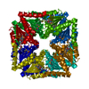 1qczS S: Starting model for refinement |
|---|---|
| Similar structure data | |
| Other databases |


-Links
 PDBj
PDBj- Assembly
| Deposited unit | 
| ||||||||
|---|---|---|---|---|---|---|---|---|---|
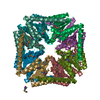
| 1 | x 8
| ||||||||
| Unit cell |
| ||||||||
| Components on special symmetry positions |
|
-Components
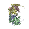
| #1: Protein | Mass: 20070.227 Da / Num. of mol.: 1 Source method: isolated from a genetically manipulated source Source: (gene. exp.) Thermotoga maritima (bacteria) / Gene: PURE OR TM0446 / Production host: Escherichia coli (E. coli)References: UniProt: Q9WYS7, phosphoribosylaminoimidazole carboxylase | ||
|---|---|---|---|
| #2: Chemical | Sulfate  Mass: 96.063 Da / Num. of mol.: 2 / Source method: obtained synthetically / Formula: SO4 Mass: 96.063 Da / Num. of mol.: 2 / Source method: obtained synthetically / Formula: SO4#3: Water | ChemComp-HOH / | Water Mass: 18.015 Da / Num. of mol.: 118 / Source method: isolated from a natural source / Formula: H2O Mass: 18.015 Da / Num. of mol.: 118 / Source method: isolated from a natural source / Formula: H2O |
-Experimental details
-Experiment
| Experiment | Method: X-RAY DIFFRACTION / Number of used crystals: 1 |
|---|
- Sample preparation
| Crystal | Density Matthews: 2.18 Å3/Da / Density % sol: 43.12 % | ||||||||||||||||||||||||||||||||||||||||||||||||||||||||
|---|---|---|---|---|---|---|---|---|---|---|---|---|---|---|---|---|---|---|---|---|---|---|---|---|---|---|---|---|---|---|---|---|---|---|---|---|---|---|---|---|---|---|---|---|---|---|---|---|---|---|---|---|---|---|---|---|---|
| Crystal grow | Temperature: 293 K / Method: vapor diffusion, sitting drop, nanodrop / pH: 5.6 Details: 2M ammonium sulfate, 0.1M tri-sodium citrate dihydrate pH 5.6, 0.2M potassium sodium tartrate tetrahydrate , VAPOR DIFFUSION, SITTING DROP, NANODROP, temperature 293K | ||||||||||||||||||||||||||||||||||||||||||||||||||||||||
| Crystal grow | *PLUS pH: 7.9 / Method: vapor diffusion | ||||||||||||||||||||||||||||||||||||||||||||||||||||||||
| Components of the solutions | *PLUS
|
-Data collection
| Diffraction | Mean temperature: 100 K |
|---|---|
| Diffraction source | Source: SYNCHROTRON / Site: SSRL  / Beamline: BL11-1 / Wavelength: 0.95369 / Beamline: BL11-1 / Wavelength: 0.95369 |
| Detector | Type: ADSC QUANTUM 315 / Detector: CCD / Date: Feb 3, 2003 / Details: flat mirror |
| Radiation | Monochromator: single crystal Si(111) bent monochromator / Protocol: SINGLE WAVELENGTH / Monochromatic (M) / Laue (L): M / Scattering type: x-ray |
| Radiation wavelength | Wavelength: 0.95369 Å / Relative weight: 1 |
| Reflection | Resolution: 1.77→24.37 Å / Num. all: 16697 / Num. obs: 16697 / % possible obs: 95.4 % / Redundancy: 7.2 % / Biso Wilson estimate: 34.3 Å2 / Rsym value: 0.072 / Net I/σ(I): 19.3 |
| Reflection shell | Resolution: 1.77→1.82 Å / Redundancy: 3.5 % / Mean I/σ(I) obs: 2.1 / Num. unique all: 931 / Rsym value: 0.452 / % possible all: 73.8 |
| Reflection | *PLUS Highest resolution: 1.77 Å / Num. obs: 16698 / Num. measured all: 119908 / Rmerge(I) obs: 0.072 |
| Reflection shell | *PLUS % possible obs: 73.8 % / Rmerge(I) obs: 0.452 / Mean I/σ(I) obs: 1.7 |
- Processing
| Software |
| |||||||||||||||||||||||||||||||||||||||||||||||||||||||||||||||||||||||||||||||||||||||||||||||||||||||||||||||||||
|---|---|---|---|---|---|---|---|---|---|---|---|---|---|---|---|---|---|---|---|---|---|---|---|---|---|---|---|---|---|---|---|---|---|---|---|---|---|---|---|---|---|---|---|---|---|---|---|---|---|---|---|---|---|---|---|---|---|---|---|---|---|---|---|---|---|---|---|---|---|---|---|---|---|---|---|---|---|---|---|---|---|---|---|---|---|---|---|---|---|---|---|---|---|---|---|---|---|---|---|---|---|---|---|---|---|---|---|---|---|---|---|---|---|---|---|---|
| Refinement | Method to determine structure: MOLECULAR REPLACEMENT Starting model: 1QCZ Resolution: 1.77→24.37 Å / Cor.coef. Fo:Fc: 0.974 / Cor.coef. Fo:Fc free: 0.964 / SU B: 4.354 / SU ML: 0.067 / TLS residual ADP flag: LIKELY RESIDUAL / Isotropic thermal model: ISOTROPIC / Cross valid method: THROUGHOUT / σ(F): 0 / ESU R: 0.103 / ESU R Free: 0.1 / Stereochemistry target values: MAXIMUM LIKELIHOOD Details: 1. WEAK, AMBIGUOUS DENSITY FOR BOTH THE N- AND C-TERMINAL RESIDUES WAS NOT MODELED. 2. UNEXPLAINED SURFACE DENSITY BETWEEN ASP11 AND ARG40, AND AT THE INTER-OCTAMER CONTACT AROUND GLU145, ...Details: 1. WEAK, AMBIGUOUS DENSITY FOR BOTH THE N- AND C-TERMINAL RESIDUES WAS NOT MODELED. 2. UNEXPLAINED SURFACE DENSITY BETWEEN ASP11 AND ARG40, AND AT THE INTER-OCTAMER CONTACT AROUND GLU145, WAS MODELED AS WATERS. 3. HYDROGENS HAVE BEEN ADDED IN THE RIDING POSITIONS
| |||||||||||||||||||||||||||||||||||||||||||||||||||||||||||||||||||||||||||||||||||||||||||||||||||||||||||||||||||
| Solvent computation | Ion probe radii: 0.8 Å / Shrinkage radii: 0.8 Å / VDW probe radii: 1.4 Å / Solvent model: BABINET MODEL WITH MASK | |||||||||||||||||||||||||||||||||||||||||||||||||||||||||||||||||||||||||||||||||||||||||||||||||||||||||||||||||||
| Displacement parameters | Biso mean: 26.807 Å2
| |||||||||||||||||||||||||||||||||||||||||||||||||||||||||||||||||||||||||||||||||||||||||||||||||||||||||||||||||||
| Refinement step | Cycle: LAST / Resolution: 1.77→24.37 Å
| |||||||||||||||||||||||||||||||||||||||||||||||||||||||||||||||||||||||||||||||||||||||||||||||||||||||||||||||||||
| Refine LS restraints |
| |||||||||||||||||||||||||||||||||||||||||||||||||||||||||||||||||||||||||||||||||||||||||||||||||||||||||||||||||||
| LS refinement shell | Resolution: 1.77→1.816 Å / Total num. of bins used: 20
| |||||||||||||||||||||||||||||||||||||||||||||||||||||||||||||||||||||||||||||||||||||||||||||||||||||||||||||||||||
| Refinement TLS params. | Method: refined / Origin x: 23.771 Å / Origin y: 1.379 Å / Origin z: 9.73 Å
| |||||||||||||||||||||||||||||||||||||||||||||||||||||||||||||||||||||||||||||||||||||||||||||||||||||||||||||||||||
| Refinement TLS group | Selection: ALL | |||||||||||||||||||||||||||||||||||||||||||||||||||||||||||||||||||||||||||||||||||||||||||||||||||||||||||||||||||
| Software | *PLUS Name: REFMAC / Version: 5.1.9999 / Classification: refinement | |||||||||||||||||||||||||||||||||||||||||||||||||||||||||||||||||||||||||||||||||||||||||||||||||||||||||||||||||||
| Refinement | *PLUS Num. reflection obs: 16697 / % reflection Rfree: 5 % / Rfactor Rfree: 0.182 / Rfactor Rwork: 0.15 | |||||||||||||||||||||||||||||||||||||||||||||||||||||||||||||||||||||||||||||||||||||||||||||||||||||||||||||||||||
| Solvent computation | *PLUS | |||||||||||||||||||||||||||||||||||||||||||||||||||||||||||||||||||||||||||||||||||||||||||||||||||||||||||||||||||
| Displacement parameters | *PLUS | |||||||||||||||||||||||||||||||||||||||||||||||||||||||||||||||||||||||||||||||||||||||||||||||||||||||||||||||||||
| Refine LS restraints | *PLUS
|
