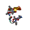
Mass: 2497.842 Da / Num. of mol.: 1 / Fragment: C-terminal domain, residues 330-348 / Mutation: all serines and threonines phosphorylated / Source method: obtained synthetically Details: This sequence occurs naturally in Bos taurus. In a synthetic peptide, all serines and threonines are phosphorylated. References: UniProt: P02699
-
Experimental details
-
Experiment
Experiment
Method: SOLUTION NMR
NMR experiment
Conditions-ID
Experiment-ID
Solution-ID
Type
1
1
1
2D TOCSY
2
2
1
2D NOESY
-
Sample preparation
Details
Contents: 0.16 mM of purified arrestin, 1.77 mM of 7PP, 7-phospho-Rh(330-348) or unphosphorylated Rh(330-348) in sodium phosphate buffer, 0.1 M, pH 6.5 and 10% D2O in a total volume of 0.6 ml Solvent system: 90% H2O/10% D2O
Method: distance geometry, constrained molecular dynamics, simulated annealing Software ordinal: 1 Details: Experimental data are for the 7-phospho-Rh(330-348) bound to arrestin. Calculations and structures submitted are without phosphate groups.
NMR representative
Selection criteria: minimized average structure
NMR ensemble
Conformer selection criteria: structures with the lowest energy Conformers calculated total number: 100 / Conformers submitted total number: 16
+
About Yorodumi
-
News
-
Feb 9, 2022. New format data for meta-information of EMDB entries
New format data for meta-information of EMDB entries
Version 3 of the EMDB header file is now the official format.
The previous official version 1.9 will be removed from the archive.
In the structure databanks used in Yorodumi, some data are registered as the other names, "COVID-19 virus" and "2019-nCoV". Here are the details of the virus and the list of structure data.
Jan 31, 2019. EMDB accession codes are about to change! (news from PDBe EMDB page)
EMDB accession codes are about to change! (news from PDBe EMDB page)
The allocation of 4 digits for EMDB accession codes will soon come to an end. Whilst these codes will remain in use, new EMDB accession codes will include an additional digit and will expand incrementally as the available range of codes is exhausted. The current 4-digit format prefixed with “EMD-” (i.e. EMD-XXXX) will advance to a 5-digit format (i.e. EMD-XXXXX), and so on. It is currently estimated that the 4-digit codes will be depleted around Spring 2019, at which point the 5-digit format will come into force.
The EM Navigator/Yorodumi systems omit the EMD- prefix.
Related info.:Q: What is EMD? / ID/Accession-code notation in Yorodumi/EM Navigator
Yorodumi is a browser for structure data from EMDB, PDB, SASBDB, etc.
This page is also the successor to EM Navigator detail page, and also detail information page/front-end page for Omokage search.
The word "yorodu" (or yorozu) is an old Japanese word meaning "ten thousand". "mi" (miru) is to see.
Related info.:EMDB / PDB / SASBDB / Comparison of 3 databanks / Yorodumi Search / Aug 31, 2016. New EM Navigator & Yorodumi / Yorodumi Papers / Jmol/JSmol / Function and homology information / Changes in new EM Navigator and Yorodumi
 Movie
Movie Controller

 Yorodumi
 Open data
 Basic information
 Components
 Keywords
 SIGNALING PROTEIN /
  Function and homology information
 Authors
 Citation
 Structure visualization
 Downloads & links
 Other downloads


 PDBj
PDBj


 Assembly
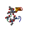
 Sample preparation
 Processing