 Movie
Movie Controller

[English] 日本語
 Yorodumi
- PDB-1nu4: U1A RNA binding domain at 1.8 angstrom resolution reveals a pre-o... -
+ Open data

- Basic information
| Entry | Database: PDB / ID: 1nu4 | ||||||
|---|---|---|---|---|---|---|---|
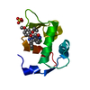
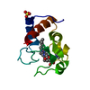
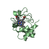
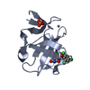
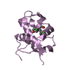
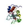
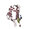
| Title | U1A RNA binding domain at 1.8 angstrom resolution reveals a pre-organized C-terminal helix | ||||||
 Components Components | U1A RNA binding domain | ||||||
 Keywords Keywords |  RNA BINDING PROTEIN / RNA recognition motif / U1 small nuclear ribonucleoprotein / RNA binding domain RNA BINDING PROTEIN / RNA recognition motif / U1 small nuclear ribonucleoprotein / RNA binding domain | ||||||
| Function / homology |  Function and homology information Function and homology informationU1 snRNP binding / U1 snRNP / U1 snRNA binding / U4/U6 x U5 tri-snRNP complex / mRNA Splicing - Major Pathway / spliceosomal complex / mRNA splicing, via spliceosome / DNA binding / RNA binding / nucleoplasm ...U1 snRNP binding / U1 snRNP / U1 snRNA binding / U4/U6 x U5 tri-snRNP complex / mRNA Splicing - Major Pathway / spliceosomal complex / mRNA splicing, via spliceosome / DNA binding / RNA binding / nucleoplasm / identical protein binding / nucleusSimilarity search - Function | ||||||
| Biological species |  Homo sapiens (human) Homo sapiens (human) | ||||||
| Method | X-RAY DIFFRACTION / MOLECULAR REPLACEMENT / Resolution: 1.8 Å | ||||||
 Authors Authors | Rupert, P.B. / Xiao, H. / Ferre-D'Amare, A.R. | ||||||
 Citation Citation | Journal: Acta Crystallogr.,Sect.D / Year: 2003 Title: U1A RNA-binding domain at 1.8 A resolution. Authors: Rupert, P.B. / Xiao, H. / Ferre-D'Amare, A.R. | ||||||
| History |
|
- Structure visualization
| Structure viewer | Molecule: MolmilJmol/JSmol |
|---|
- Downloads & links
-Download
| PDBx/mmCIF format | 1nu4.cif.gz | 58.3 KB | Display | PDBx/mmCIF format |
|---|---|---|---|---|
| PDB format | pdb1nu4.ent.gz | 42.3 KB | Display | PDB format |
| PDBx/mmJSON format | 1nu4.json.gz | Tree view | PDBx/mmJSON format | |
| Others |  Other downloads Other downloads |
-Validation report
| Arichive directory | https://data.pdbj.org/pub/pdb/validation_reports/nu/1nu4ftp://data.pdbj.org/pub/pdb/validation_reports/nu/1nu4 | HTTPS FTP |
|---|
-Related structure data
| Related structure data |  1urnS S: Starting model for refinement |
|---|---|
| Similar structure data |

-Links
 PDBj
PDBj


- Assembly
| Deposited unit | 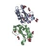
| |||||||||||||||||||||||||||||||||
|---|---|---|---|---|---|---|---|---|---|---|---|---|---|---|---|---|---|---|---|---|---|---|---|---|---|---|---|---|---|---|---|---|---|---|
| 1 | 
| |||||||||||||||||||||||||||||||||
| 2 | 
| |||||||||||||||||||||||||||||||||
| 3 | 
| |||||||||||||||||||||||||||||||||
| 4 | 
| |||||||||||||||||||||||||||||||||
| 5 | 
| |||||||||||||||||||||||||||||||||
| 6 | 
| |||||||||||||||||||||||||||||||||
| 7 | 
| |||||||||||||||||||||||||||||||||
| Unit cell |
| |||||||||||||||||||||||||||||||||
| Components on special symmetry positions |
|
-Components
| #1: Protein | Mass: 11209.120 Da / Num. of mol.: 2 / Fragment: U1A RBD1 / Mutation: Y31H,Q36R Source method: isolated from a genetically manipulated source Source: (gene. exp.) Homo sapiens (human) / Production host:  Escherichia coli (E. coli) / References: UniProt: P09012 Escherichia coli (E. coli) / References: UniProt: P09012#2: Chemical | ChemComp-MG /   Mass: 24.305 Da / Num. of mol.: 5 / Source method: obtained synthetically / Formula: Mg Mass: 24.305 Da / Num. of mol.: 5 / Source method: obtained synthetically / Formula: Mg#3: Chemical | ChemComp-MLA / Malonic acid  Mass: 104.061 Da / Num. of mol.: 12 / Source method: obtained synthetically / Formula: C3H4O4 Mass: 104.061 Da / Num. of mol.: 12 / Source method: obtained synthetically / Formula: C3H4O4#4: Water | ChemComp-HOH / | Water Mass: 18.015 Da / Num. of mol.: 99 / Source method: isolated from a natural source / Formula: H2O Mass: 18.015 Da / Num. of mol.: 99 / Source method: isolated from a natural source / Formula: H2O |
|---|
-Experimental details
-Experiment
| Experiment | Method: X-RAY DIFFRACTION / Number of used crystals: 1 |
|---|
- Sample preparation
| Crystal | Density Matthews: 2.18 Å3/Da / Density % sol: 43.56 % | |||||||||||||||||||||||||||||||||||||||||||||||||
|---|---|---|---|---|---|---|---|---|---|---|---|---|---|---|---|---|---|---|---|---|---|---|---|---|---|---|---|---|---|---|---|---|---|---|---|---|---|---|---|---|---|---|---|---|---|---|---|---|---|---|
| Crystal grow | Temperature: 298 K / Method: vapor diffusion, hanging drop / pH: 5 Details: Potassium Malonate, pH 5.0, VAPOR DIFFUSION, HANGING DROP, temperature 298K | |||||||||||||||||||||||||||||||||||||||||||||||||
| Crystal grow | *PLUS pH: 6.5 | |||||||||||||||||||||||||||||||||||||||||||||||||
| Components of the solutions | *PLUS
|
-Data collection
| Diffraction | Mean temperature: 100 K |
|---|---|
| Diffraction source | Source: ROTATING ANODE / Type: RIGAKU RU300 / Wavelength: 1.5418 Å |
| Detector | Type: RIGAKU RAXIS IV / Detector: IMAGE PLATE / Date: Aug 15, 2002 / Details: osmics mirrors |
| Radiation | Monochromator: graphite / Protocol: SINGLE WAVELENGTH / Monochromatic (M) / Laue (L): M / Scattering type: x-ray |
| Radiation wavelength | Wavelength: 1.5418 Å / Relative weight: 1 |
| Reflection | Resolution: 1.8→30 Å / Num. all: 18308 / Num. obs: 17618 / % possible obs: 96.2 % / Observed criterion σ(F): 0 / Observed criterion σ(I): 0 / Redundancy: 4 % / Biso Wilson estimate: 31.1 Å2 / Rmerge(I) obs: 0.031 / Rsym value: 0.031 / Net I/σ(I): 37.6 |
| Reflection shell | Resolution: 1.8→1.86 Å / Redundancy: 1.4 % / Rmerge(I) obs: 0.311 / Mean I/σ(I) obs: 2.46 / Num. unique all: 1365 / Rsym value: 0.311 / % possible all: 76.8 |
| Reflection | *PLUS Lowest resolution: 30 Å / Num. measured all: 70351 |
| Reflection shell | *PLUS % possible obs: 76.8 % |
- Processing
| Software |
| ||||||||||||||||||||||||||||||||||||
|---|---|---|---|---|---|---|---|---|---|---|---|---|---|---|---|---|---|---|---|---|---|---|---|---|---|---|---|---|---|---|---|---|---|---|---|---|---|
| Refinement | Method to determine structure: MOLECULAR REPLACEMENT Starting model: pdb entry 1urn Resolution: 1.8→29.16 Å / Rfactor Rfree error: 0.006 / Isotropic thermal model: RESTRAINED / Cross valid method: THROUGHOUT / σ(F): 0 / σ(I): 0 / Stereochemistry target values: Engh & Huber
| ||||||||||||||||||||||||||||||||||||
| Solvent computation | Solvent model: FLAT MODEL / Bsol: 57.9717 Å2 / ksol: 0.401443 e/Å3 | ||||||||||||||||||||||||||||||||||||
| Displacement parameters | Biso mean: 40.3 Å2
| ||||||||||||||||||||||||||||||||||||
| Refine analyze |
| ||||||||||||||||||||||||||||||||||||
| Refinement step | Cycle: LAST / Resolution: 1.8→29.16 Å
| ||||||||||||||||||||||||||||||||||||
| Refine LS restraints |
| ||||||||||||||||||||||||||||||||||||
| LS refinement shell | Resolution: 1.8→1.91 Å / Rfactor Rfree error: 0.023 / Total num. of bins used: 6
| ||||||||||||||||||||||||||||||||||||
| Xplor file |
| ||||||||||||||||||||||||||||||||||||
| Refinement | *PLUS Highest resolution: 1.8 Å / Lowest resolution: 30 Å / % reflection Rfree: 10 % / Rfactor Rfree: 0.2344 / Rfactor Rwork: 0.2032 | ||||||||||||||||||||||||||||||||||||
| Solvent computation | *PLUS | ||||||||||||||||||||||||||||||||||||
| Displacement parameters | *PLUS | ||||||||||||||||||||||||||||||||||||
| Refine LS restraints | *PLUS
|
