 Movie
Movie Controller

[English] 日本語
 Yorodumi
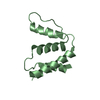
- PDB-1nti: RDC-refined NMR structure of bovine Acyl-coenzyme A Binding Prote... -
+ Open data

- Basic information
| Entry | Database: PDB / ID: 1nti | ||||||
|---|---|---|---|---|---|---|---|
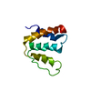
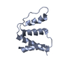
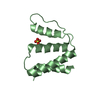
| Title | RDC-refined NMR structure of bovine Acyl-coenzyme A Binding Protein, ACBP | ||||||
 Components Components | Acyl-CoA-binding protein | ||||||
 Keywords Keywords | LIPID BINDING PROTEIN / four-helix-bundle / ACBP family | ||||||
| Function / homology |  Function and homology information Function and homology informationMitochondrial Fatty Acid Beta-Oxidation / fatty-acyl-CoA binding / fatty acid metabolic process / Golgi apparatus / endoplasmic reticulumSimilarity search - Function | ||||||
| Biological species |  Bos taurus (cattle) Bos taurus (cattle) | ||||||
| Method | SOLUTION NMR / simulated annealing, molecular dynamics | ||||||
 Authors Authors | Lerche, M.H. / Kragelund, B.B. / Redfield, C. / Poulsen, F.M. | ||||||
 Citation Citation | Journal: To be Published Title: Subtle structural response to ligand binding revealed by residual dipolar coupling refined NMR structures of acyl coenzyme A binding protein Authors: Lerche, M.H. / Kragelund, B.B. / Redfield, C. / Poulsen, F.M. #1: Journal: J.Biomol.NMR / Year: 1993Title: The three-dimensional structure of acyl-coenzyme A binding protein from bovine liver: structural refinement using heteronuclear multidimensional NMR spectroscopy. Authors: Andersen, K.V. / Poulsen, F.M. #2: Journal: J.Mol.Biol. / Year: 1993Title: Three-dimensional structure of the complex between acyl-coenzyme A binding protein and palmitoyl-coenzyme A. Authors: Kragelund, B.B. / Andersen, K.V. / Madsen, J.C. / Knudsen, J. / Poulsen, F.M. | ||||||
| History |
|
- Structure visualization
| Structure viewer | Molecule: MolmilJmol/JSmol |
|---|
- Downloads & links
-Download
| PDBx/mmCIF format | 1nti.cif.gz | 598.8 KB | Display | PDBx/mmCIF format |
|---|---|---|---|---|
| PDB format | pdb1nti.ent.gz | 512.6 KB | Display | PDB format |
| PDBx/mmJSON format | 1nti.json.gz | Tree view | PDBx/mmJSON format | |
| Others |  Other downloads Other downloads |
-Validation report
| Arichive directory | https://data.pdbj.org/pub/pdb/validation_reports/nt/1ntiftp://data.pdbj.org/pub/pdb/validation_reports/nt/1nti | HTTPS FTP |
|---|
-Related structure data
| Related structure data | |
|---|---|
| Similar structure data |


-Links
 PDBj
PDBj
- Assembly
| Deposited unit | 
| |||||||||
|---|---|---|---|---|---|---|---|---|---|---|
| 1 |
| |||||||||
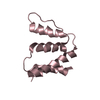
| NMR ensembles |
|
-Components
| #1: Protein | / ACBP Mass: 9931.289 Da / Num. of mol.: 1 Source method: isolated from a genetically manipulated source Source: (gene. exp.) Bos taurus (cattle) / Production host:  Escherichia coli (E. coli) / References: UniProt: P07107 Escherichia coli (E. coli) / References: UniProt: P07107 |
|---|
-Experimental details
-Experiment
| Experiment | Method: SOLUTION NMR | ||||||||||||
|---|---|---|---|---|---|---|---|---|---|---|---|---|---|
| NMR experiment |
| ||||||||||||
| NMR details | Text: refinement of earlier pdb deposit, 2ABD, see entry for detail. NOE and dihedral angle constraints obtained from this entry. Residual dipolar couplings added as additional restraints |
- Sample preparation
| Details |
| |||||||||
|---|---|---|---|---|---|---|---|---|---|---|
| Sample conditions | Ionic strength: no salt added / pH: 6.5 / Pressure: ambient / Temperature: 310 K |
-NMR measurement
| Radiation | Protocol: SINGLE WAVELENGTH / Monochromatic (M) / Laue (L): M | |||||||||||||||
|---|---|---|---|---|---|---|---|---|---|---|---|---|---|---|---|---|
| Radiation wavelength | Relative weight: 1 | |||||||||||||||
| NMR spectrometer |
|
- Processing
| NMR software | Name: X-PLOR / Version: modified 3.8 / Classification: refinement |
|---|---|
| Refinement | Method: simulated annealing, molecular dynamics / Software ordinal: 1 |
| NMR representative | Selection criteria: lowest energy |
| NMR ensemble | Conformer selection criteria: structures with the lowest energy Conformers calculated total number: 100 / Conformers submitted total number: 20 |
