 Movie
Movie Controller

[English] 日本語
 Yorodumi
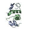
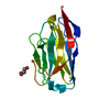
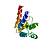
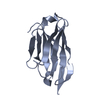
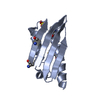
- PDB-1nqj: CRYSTAL STRUCTURE OF CLOSTRIDIUM HISTOLYTICUM COLG COLLAGENASE CO... -
+ Open data

- Basic information
| Entry | Database: PDB / ID: 1nqj | ||||||
|---|---|---|---|---|---|---|---|
| Title | CRYSTAL STRUCTURE OF CLOSTRIDIUM HISTOLYTICUM COLG COLLAGENASE COLLAGEN-BINDING DOMAIN 3B AT 1.0 ANGSTROM RESOLUTION IN ABSENCE OF CALCIUM | ||||||
 Components Components | class 1 collagenase | ||||||
 Keywords Keywords |  HYDROLASE / BETA SANDWICH / METALLOPROTEASE / COLLAGEN-BINDING DOMAIN / LITHIUM / CHLORINE HYDROLASE / BETA SANDWICH / METALLOPROTEASE / COLLAGEN-BINDING DOMAIN / LITHIUM / CHLORINE | ||||||
| Function / homology |  Function and homology informationtripeptidase activity / microbial collagenase / collagen metabolic process / collagen binding / metalloendopeptidase activity / endopeptidase activity / calcium ion binding / proteolysis / zinc ion binding / extracellular region Function and homology informationtripeptidase activity / microbial collagenase / collagen metabolic process / collagen binding / metalloendopeptidase activity / endopeptidase activity / calcium ion binding / proteolysis / zinc ion binding / extracellular regionSimilarity search - Function | ||||||
| Biological species |  Clostridium histolyticum (bacteria) Clostridium histolyticum (bacteria) | ||||||
| Method | X-RAY DIFFRACTION / SYNCHROTRON / MAD / Resolution: 1 Å | ||||||
 Authors Authors | Wilson, J.J. / Matsushita, O. / Okabe, A. / Sakon, J. | ||||||
 Citation Citation | Journal: Embo J. / Year: 2003 Title: A bacterial collagen-binding domain with novel calcium-binding motif controls domain orientation Authors: Wilson, J.J. / Matsushita, O. / Okabe, A. / Sakon, J. | ||||||
| History |
|
- Structure visualization
| Structure viewer | Molecule: MolmilJmol/JSmol |
|---|
- Downloads & links
-Download
| PDBx/mmCIF format | 1nqj.cif.gz | 163 KB | Display | PDBx/mmCIF format |
|---|---|---|---|---|
| PDB format | pdb1nqj.ent.gz | 130.5 KB | Display | PDB format |
| PDBx/mmJSON format | 1nqj.json.gz | Tree view | PDBx/mmJSON format | |
| Others |  Other downloads Other downloads |
-Validation report
| Arichive directory | https://data.pdbj.org/pub/pdb/validation_reports/nq/1nqjftp://data.pdbj.org/pub/pdb/validation_reports/nq/1nqj | HTTPS FTP |
|---|
-Related structure data


-Links
 PDBj
PDBj
- Assembly
| Deposited unit | 
| ||||||||
|---|---|---|---|---|---|---|---|---|---|
| 1 | 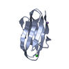
| ||||||||
| 2 | 
| ||||||||
| Unit cell |
| ||||||||
| Details | The biological assembly is a monomer. |
-Components
| #1: Protein | Mass: 13507.840 Da / Num. of mol.: 2 / Fragment: COLLAGEN-BINDING DOMAIN Source method: isolated from a genetically manipulated source Source: (gene. exp.) Clostridium histolyticum (bacteria) / Gene: ColG / Plasmid: pGEX-4T-2 / Species (production host): Escherichia coli / Production host: Escherichia coli BL21 (bacteria) / Strain (production host): BL21References: UniProt: Q9S0X0, UniProt: Q9X721*PLUS, microbial collagenase#2: Chemical | Chloride  Mass: 35.453 Da / Num. of mol.: 2 / Source method: obtained synthetically / Formula: Cl Mass: 35.453 Da / Num. of mol.: 2 / Source method: obtained synthetically / Formula: Cl#3: Chemical | Lithium  Mass: 6.941 Da / Num. of mol.: 3 / Source method: obtained synthetically / Formula: Li Mass: 6.941 Da / Num. of mol.: 3 / Source method: obtained synthetically / Formula: Li#4: Water | ChemComp-HOH / | Water Mass: 18.015 Da / Num. of mol.: 388 / Source method: isolated from a natural source / Formula: H2O Mass: 18.015 Da / Num. of mol.: 388 / Source method: isolated from a natural source / Formula: H2O |
|---|
-Experimental details
-Experiment
| Experiment | Method: X-RAY DIFFRACTION / Number of used crystals: 1 |
|---|
- Sample preparation
| Crystal | Density Matthews: 1.71 Å3/Da / Density % sol: 27.71 % | |||||||||||||||||||||||||||||||||||||||||||||||||
|---|---|---|---|---|---|---|---|---|---|---|---|---|---|---|---|---|---|---|---|---|---|---|---|---|---|---|---|---|---|---|---|---|---|---|---|---|---|---|---|---|---|---|---|---|---|---|---|---|---|---|
| Crystal grow | Temperature: 277 K / Method: vapor diffusion, hanging drop / pH: 4.6 Details: PEG 3350, LITHIUM CHLORIDE, SODIUM ACETATE, pH 4.6, VAPOR DIFFUSION, HANGING DROP, temperature 277K | |||||||||||||||||||||||||||||||||||||||||||||||||
| Crystal grow | *PLUS Temperature: 4 ℃ | |||||||||||||||||||||||||||||||||||||||||||||||||
| Components of the solutions | *PLUS
|
-Data collection
| Diffraction | Mean temperature: 100 K |
|---|---|
| Diffraction source | Source: SYNCHROTRON / Site: APS  / Beamline: 17-ID / Wavelength: 1 / Wavelength: 1 Å / Beamline: 17-ID / Wavelength: 1 / Wavelength: 1 Å |
| Detector | Type: MARRESEARCH / Detector: CCD / Date: Jun 1, 2000 |
| Radiation | Monochromator: SI (111) / Protocol: SINGLE WAVELENGTH / Monochromatic (M) / Laue (L): M / Scattering type: x-ray |
| Radiation wavelength | Wavelength: 1 Å / Relative weight: 1 |
| Reflection | Resolution: 0.983→36 Å / Num. all: 114718 / Num. obs: 105185 / % possible obs: 91.7 % / Observed criterion σ(F): 0 / Observed criterion σ(I): 0 / Redundancy: 3.01 % / Rsym value: 0.075 / Net I/σ(I): 30.7 |
| Reflection shell | Resolution: 1→1.07 Å / Mean I/σ(I) obs: 2.94 / Num. unique all: 15310 / Rsym value: 0.318 / % possible all: 72.64 |
| Reflection | *PLUS Highest resolution: 1 Å / Lowest resolution: 36 Å / Rmerge(I) obs: 0.074 |
| Reflection shell | *PLUS % possible obs: 72.64 % |
- Processing
| Software |
| |||||||||||||||||||||||||||||||||||
|---|---|---|---|---|---|---|---|---|---|---|---|---|---|---|---|---|---|---|---|---|---|---|---|---|---|---|---|---|---|---|---|---|---|---|---|---|
| Refinement | Method to determine structure: MAD / Resolution: 1→28.85 Å / Num. parameters: 20305 / Num. restraintsaints: 37831 / Cross valid method: FREE R / σ(F): 0 / Stereochemistry target values: Engh & Huber
| |||||||||||||||||||||||||||||||||||
| Refine analyze | Num. disordered residues: 40 / Occupancy sum hydrogen: 1650 / Occupancy sum non hydrogen: 2066.53 | |||||||||||||||||||||||||||||||||||
| Refinement step | Cycle: LAST / Resolution: 1→28.85 Å
| |||||||||||||||||||||||||||||||||||
| Refine LS restraints |
| |||||||||||||||||||||||||||||||||||
| LS refinement shell |
| |||||||||||||||||||||||||||||||||||
| Software | *PLUS Name: SHELXL / Version: 97 / Classification: refinement | |||||||||||||||||||||||||||||||||||
| Refinement | *PLUS Lowest resolution: 28.9 Å / % reflection Rfree: 10 % / Rfactor Rfree: 0.166 / Rfactor Rwork: 0.142 | |||||||||||||||||||||||||||||||||||
| Solvent computation | *PLUS | |||||||||||||||||||||||||||||||||||
| Displacement parameters | *PLUS | |||||||||||||||||||||||||||||||||||
| Refine LS restraints | *PLUS
|
