 Movie
Movie Controller

[English] 日本語
 Yorodumi
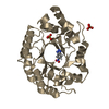
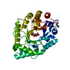
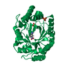
- PDB-1nq6: Crystal Structure of the catalytic domain of xylanase A from Stre... -
+ Open data

- Basic information
| Entry | Database: PDB / ID: 1nq6 | ||||||
|---|---|---|---|---|---|---|---|
| Title | Crystal Structure of the catalytic domain of xylanase A from Streptomyces halstedii JM8 | ||||||
 Components Components | Xys1 | ||||||
 Keywords Keywords |  HYDROLASE / GLYCOSIDE HYDROLASE FAMILY 10 / XYLANASE / XYLAN DEGRADATION HYDROLASE / GLYCOSIDE HYDROLASE FAMILY 10 / XYLANASE / XYLAN DEGRADATION | ||||||
| Function / homology |  Function and homology informationpolysaccharide binding / endo-1,4-beta-xylanase activity / endo-1,4-beta-xylanase / polysaccharide catabolic process Function and homology informationpolysaccharide binding / endo-1,4-beta-xylanase activity / endo-1,4-beta-xylanase / polysaccharide catabolic processSimilarity search - Function | ||||||
| Biological species |  Streptomyces halstedii (bacteria) Streptomyces halstedii (bacteria) | ||||||
| Method | X-RAY DIFFRACTION / SYNCHROTRON / MOLECULAR REPLACEMENT / Resolution: 1.78 Å | ||||||
 Authors Authors | Canals, A. / Vega, M.C. / Gomis-Ruth, F.X. / Santamaria, R.I. / Coll, M. | ||||||
 Citation Citation | Journal: Acta Crystallogr.,Sect.D / Year: 2003 Title: Structure of xylanase Xys1delta from Streptomyces halstedii. Authors: Canals, A. / Vega, M.C. / Gomis-Ruth, F.X. / Diaz, M. / Santamaria R, R.I. / Coll, M. | ||||||
| History |
|
- Structure visualization
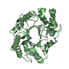
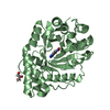
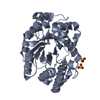
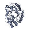
| Structure viewer | Molecule: MolmilJmol/JSmol |
|---|
- Downloads & links
-Download
| PDBx/mmCIF format | 1nq6.cif.gz | 78.3 KB | Display | PDBx/mmCIF format |
|---|---|---|---|---|
| PDB format | pdb1nq6.ent.gz | 56.7 KB | Display | PDB format |
| PDBx/mmJSON format | 1nq6.json.gz | Tree view | PDBx/mmJSON format | |
| Others |  Other downloads Other downloads |
-Validation report
| Arichive directory | https://data.pdbj.org/pub/pdb/validation_reports/nq/1nq6ftp://data.pdbj.org/pub/pdb/validation_reports/nq/1nq6 | HTTPS FTP |
|---|
-Related structure data
| Related structure data |  1e0wS S: Starting model for refinement |
|---|---|
| Similar structure data |


-Links
 PDBj
PDBj

- Assembly
| Deposited unit | 
| ||||||||
|---|---|---|---|---|---|---|---|---|---|
| 1 |
| ||||||||
| Unit cell |
|
-Components
| #1: Protein | Mass: 32614.070 Da / Num. of mol.: 1 / Fragment: Catalytic domain Source method: isolated from a genetically manipulated source Source: (gene. exp.) Streptomyces halstedii (bacteria) / Strain: JM8 / Gene: xysA / Plasmid: pJM9 / Production host: Streptomyces parvulus (bacteria) / Strain (production host): JI2283 / References: UniProt: Q59922, endo-1,4-beta-xylanase |
|---|---|
| #2: Chemical | ChemComp-MG /   Mass: 24.305 Da / Num. of mol.: 1 / Source method: obtained synthetically / Formula: Mg Mass: 24.305 Da / Num. of mol.: 1 / Source method: obtained synthetically / Formula: Mg |
| #3: Water | ChemComp-HOH / Water Mass: 18.015 Da / Num. of mol.: 341 / Source method: isolated from a natural source / Formula: H2O Mass: 18.015 Da / Num. of mol.: 341 / Source method: isolated from a natural source / Formula: H2O |
-Experimental details
-Experiment
| Experiment | Method: X-RAY DIFFRACTION / Number of used crystals: 1 |
|---|
- Sample preparation
| Crystal | Density Matthews: 1.61 Å3/Da / Density % sol: 22.93 % | |||||||||||||||||||||||||||||||||||
|---|---|---|---|---|---|---|---|---|---|---|---|---|---|---|---|---|---|---|---|---|---|---|---|---|---|---|---|---|---|---|---|---|---|---|---|---|
| Crystal grow | Temperature: 298 K / Method: vapor diffusion, hanging drop / pH: 5.5 Details: PEG 4000, MAGNESIUM CHLORIDE, SODIUM ACETATE, pH 5.5, VAPOR DIFFUSION, HANGING DROP, temperature 298K | |||||||||||||||||||||||||||||||||||
| Crystal grow | *PLUS Method: vapor diffusion, hanging drop | |||||||||||||||||||||||||||||||||||
| Components of the solutions | *PLUS
|
-Data collection
| Diffraction | Mean temperature: 120 K |
|---|---|
| Diffraction source | Source: SYNCHROTRON / Site: EMBL/DESY, HAMBURG  / Beamline: BW7B / Wavelength: 0.885 Å / Beamline: BW7B / Wavelength: 0.885 Å |
| Detector | Type: MARRESEARCH / Detector: IMAGE PLATE / Date: Jul 22, 1996 |
| Radiation | Monochromator: MIRRORS / Protocol: SINGLE WAVELENGTH / Monochromatic (M) / Laue (L): M / Scattering type: x-ray |
| Radiation wavelength | Wavelength: 0.885 Å / Relative weight: 1 |
| Reflection | Resolution: 1.782→24.818 Å / Num. all: 23589 / Num. obs: 22905 / % possible obs: 97.1 % / Observed criterion σ(F): 0 / Observed criterion σ(I): 0 / Biso Wilson estimate: 9.453 Å2 / Rmerge(I) obs: 0.044 / Net I/σ(I): 13.1 |
| Reflection shell | Resolution: 1.78→1.88 Å / Rmerge(I) obs: 0.084 / Mean I/σ(I) obs: 8.9 / % possible all: 85.8 |
| Reflection | *PLUS Highest resolution: 1.78 Å / Lowest resolution: 25 Å / Redundancy: 3.4 % / Num. measured all: 78251 |
| Reflection shell | *PLUS % possible obs: 85.8 % / Redundancy: 2.7 % |
- Processing
| Software |
| |||||||||||||||||||||||||||||||||||||||||||||||||||||||||||||||||||||||||||
|---|---|---|---|---|---|---|---|---|---|---|---|---|---|---|---|---|---|---|---|---|---|---|---|---|---|---|---|---|---|---|---|---|---|---|---|---|---|---|---|---|---|---|---|---|---|---|---|---|---|---|---|---|---|---|---|---|---|---|---|---|---|---|---|---|---|---|---|---|---|---|---|---|---|---|---|---|
| Refinement | Method to determine structure: MOLECULAR REPLACEMENT Starting model: PDB ENTRY 1E0W Resolution: 1.78→24.82 Å / Cor.coef. Fo:Fc: 0.96 / Cor.coef. Fo:Fc free: 0.941 / SU B: 2.269 / SU ML: 0.072 / TLS residual ADP flag: LIKELY RESIDUAL / Cross valid method: THROUGHOUT / σ(F): 0 / σ(I): 0 / ESU R: 0.134 / ESU R Free: 0.115 / Stereochemistry target values: MAXIMUM LIKELIHOOD
| |||||||||||||||||||||||||||||||||||||||||||||||||||||||||||||||||||||||||||
| Solvent computation | Ion probe radii: 0.8 Å / Shrinkage radii: 0.8 Å / VDW probe radii: 1.4 Å / Solvent model: BABINET MODEL WITH MASK | |||||||||||||||||||||||||||||||||||||||||||||||||||||||||||||||||||||||||||
| Displacement parameters | Biso mean: 13.257 Å2
| |||||||||||||||||||||||||||||||||||||||||||||||||||||||||||||||||||||||||||
| Refinement step | Cycle: LAST / Resolution: 1.78→24.82 Å
| |||||||||||||||||||||||||||||||||||||||||||||||||||||||||||||||||||||||||||
| Refine LS restraints |
| |||||||||||||||||||||||||||||||||||||||||||||||||||||||||||||||||||||||||||
| LS refinement shell | Resolution: 1.78→1.826 Å / Total num. of bins used: 20
| |||||||||||||||||||||||||||||||||||||||||||||||||||||||||||||||||||||||||||
| Refinement TLS params. | Method: refined / Refine-ID: X-RAY DIFFRACTION
| |||||||||||||||||||||||||||||||||||||||||||||||||||||||||||||||||||||||||||
| Refinement TLS group |
| |||||||||||||||||||||||||||||||||||||||||||||||||||||||||||||||||||||||||||
| Refinement | *PLUS Lowest resolution: 25 Å / Rfactor Rfree: 0.18 / Rfactor Rwork: 0.15 | |||||||||||||||||||||||||||||||||||||||||||||||||||||||||||||||||||||||||||
| Solvent computation | *PLUS | |||||||||||||||||||||||||||||||||||||||||||||||||||||||||||||||||||||||||||
| Displacement parameters | *PLUS | |||||||||||||||||||||||||||||||||||||||||||||||||||||||||||||||||||||||||||
| Refine LS restraints | *PLUS
| |||||||||||||||||||||||||||||||||||||||||||||||||||||||||||||||||||||||||||
| LS refinement shell | *PLUS Rfactor Rfree: 0.22 / Rfactor Rwork: 0.16 |
