 Movie
Movie Controller

[English] 日本語
 Yorodumi
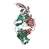
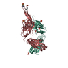
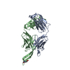
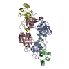
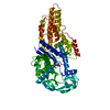
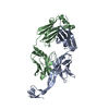
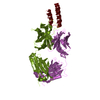
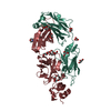
- PDB-1nl0: Crystal structure of human factor IX Gla domain in complex of an ... -
+ Open data

- Basic information
| Entry | Database: PDB / ID: 1nl0 | ||||||
|---|---|---|---|---|---|---|---|
| Title | Crystal structure of human factor IX Gla domain in complex of an inhibitory antibody, 10C12 | ||||||
 Components Components |
| ||||||
 Keywords Keywords |  IMMUNE SYSTEM / ANTIBODY / GLA DOMAIN IMMUNE SYSTEM / ANTIBODY / GLA DOMAIN | ||||||
| Function / homology |  Function and homology information Function and homology informationDefective F9 secretion / Defective gamma-carboxylation of F9 / coagulation factor IXa / Defective F9 activation / Defective factor IX causes thrombophilia / Defective cofactor function of FVIIIa variant / Defective F9 variant does not activate FX / zymogen activation / Extrinsic Pathway of Fibrin Clot Formation / Protein hydroxylation ...Defective F9 secretion / Defective gamma-carboxylation of F9 / coagulation factor IXa / Defective F9 activation / Defective factor IX causes thrombophilia / Defective cofactor function of FVIIIa variant / Defective F9 variant does not activate FX / zymogen activation / Extrinsic Pathway of Fibrin Clot Formation / Protein hydroxylation / Gamma-carboxylation of protein precursors / Transport of gamma-carboxylated protein precursors from the endoplasmic reticulum to the Golgi apparatus / Removal of aminoterminal propeptides from gamma-carboxylated proteins / Intrinsic Pathway of Fibrin Clot Formation / Golgi lumen / blood coagulation / collagen-containing extracellular matrix / endopeptidase activity / endoplasmic reticulum lumen / serine-type endopeptidase activity / calcium ion binding / proteolysis / extracellular space / extracellular exosome / extracellular region / metal ion binding / plasma membraneSimilarity search - Function | ||||||
| Biological species |  Homo sapiens (human) Homo sapiens (human) | ||||||
| Method | X-RAY DIFFRACTION / SYNCHROTRON / MOLECULAR REPLACEMENT / Resolution: 2.2 Å | ||||||
 Authors Authors | Huang, M. / Furie, B.C. / Furie, B. | ||||||
 Citation Citation | Journal: J.Biol.Chem. / Year: 2004 Title: Crystal Structure of the Calcium-stabilized Human Factor IX Gla Domain Bound to a Conformation-specific Anti-factor IX Antibody. Authors: Huang, M. / Furie, B.C. / Furie, B. | ||||||
| History |
| ||||||
| Remark 999 | SEQUENCE an appropriate sequence database reference was not available for chains L and H at the ...SEQUENCE an appropriate sequence database reference was not available for chains L and H at the time of processing. |
- Structure visualization
| Structure viewer | Molecule: MolmilJmol/JSmol |
|---|
- Downloads & links
-Download
| PDBx/mmCIF format | 1nl0.cif.gz | 110.3 KB | Display | PDBx/mmCIF format |
|---|---|---|---|---|
| PDB format | pdb1nl0.ent.gz | 83.6 KB | Display | PDB format |
| PDBx/mmJSON format | 1nl0.json.gz | Tree view | PDBx/mmJSON format | |
| Others |  Other downloads Other downloads |
-Validation report
| Arichive directory | https://data.pdbj.org/pub/pdb/validation_reports/nl/1nl0ftp://data.pdbj.org/pub/pdb/validation_reports/nl/1nl0 | HTTPS FTP |
|---|
-Related structure data


-Links
 PDBj
PDBj


- Assembly
| Deposited unit | 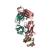
| ||||||||
|---|---|---|---|---|---|---|---|---|---|
| 1 |
| ||||||||
| 2 | 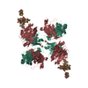
| ||||||||
| Unit cell |
| ||||||||
| Components on special symmetry positions |
|
-Components
-Protein , 1 types, 1 molecules G
| #3: Protein | Mass: 7117.892 Da / Num. of mol.: 1 / Fragment: Gla domain / Source method: obtained synthetically Details: The protein was chemically synthesized. THE SEQUENCE OF THE PROTEIN IS NATURALLY FOUND IN HOMO SAPIENS. References: UniProt: P00740 |
|---|
-Antibody , 2 types, 2 molecules LH
| #1: Antibody | Mass: 22631.020 Da / Num. of mol.: 1 Source method: isolated from a genetically manipulated source Source: (gene. exp.) Homo sapiens (human) / Gene: 10C12 / Production host:  Escherichia coli (E. coli) Escherichia coli (E. coli) |
|---|---|
| #2: Antibody | Mass: 23800.863 Da / Num. of mol.: 1 Source method: isolated from a genetically manipulated source Source: (gene. exp.) Homo sapiens (human) / Gene: 10C12 / Production host: Escherichia coli (E. coli) |
-Non-polymers , 3 types, 155 molecules 


| #4: Chemical | ChemComp-SO4 / Sulfate Mass: 96.063 Da / Num. of mol.: 1 / Source method: obtained synthetically / Formula: SO4 Mass: 96.063 Da / Num. of mol.: 1 / Source method: obtained synthetically / Formula: SO4 | ||
|---|---|---|---|
| #5: Chemical | ChemComp-CA /  Mass: 40.078 Da / Num. of mol.: 6 / Source method: obtained synthetically / Formula: Ca Mass: 40.078 Da / Num. of mol.: 6 / Source method: obtained synthetically / Formula: Ca#6: Water | ChemComp-HOH / | WaterMass: 18.015 Da / Num. of mol.: 148 / Source method: isolated from a natural source / Formula: H2O |
-Experimental details
-Experiment
| Experiment | Method: X-RAY DIFFRACTION / Number of used crystals: 1 |
|---|
- Sample preparation
| Crystal | Density Matthews: 2.79 Å3/Da / Density % sol: 55.87 % | |||||||||||||||||||||||||||||||||||||||||||||||||||||||||||||||
|---|---|---|---|---|---|---|---|---|---|---|---|---|---|---|---|---|---|---|---|---|---|---|---|---|---|---|---|---|---|---|---|---|---|---|---|---|---|---|---|---|---|---|---|---|---|---|---|---|---|---|---|---|---|---|---|---|---|---|---|---|---|---|---|---|
| Crystal grow | *PLUS pH: 7.5 / Method: vapor diffusion, sitting drop | |||||||||||||||||||||||||||||||||||||||||||||||||||||||||||||||
| Components of the solutions | *PLUS
|
-Data collection
| Diffraction | Mean temperature: 103 K |
|---|---|
| Diffraction source | Source: SYNCHROTRON / Site: NSLS  / Beamline: X25 / Wavelength: 1.08 Å / Beamline: X25 / Wavelength: 1.08 Å |
| Detector | Type: ADSC QUANTUM 4 / Detector: CCD |
| Radiation | Protocol: SINGLE WAVELENGTH / Monochromatic (M) / Laue (L): M / Scattering type: x-ray |
| Radiation wavelength | Wavelength: 1.08 Å / Relative weight: 1 |
| Reflection | Resolution: 2.1→24.66 Å / Num. obs: 28585 / % possible obs: 81.9 % / Observed criterion σ(I): -3 / Redundancy: 8.4 % / Biso Wilson estimate: 23.3 Å2 / Rmerge(I) obs: 0.05 / Net I/σ(I): 25.7 |
| Reflection shell | Resolution: 2.1→2.18 Å / Rmerge(I) obs: 0.119 / Mean I/σ(I) obs: 5.4 / % possible all: 38.7 |
| Reflection | *PLUS Num. measured all: 241036 / Rmerge(I) obs: 0.05 |
| Reflection shell | *PLUS % possible obs: 38.7 % / Num. unique obs: 1353 |
- Processing
| Software |
| ||||||||||||||||||||||||||||||||||||||||||||||||||||||||||||
|---|---|---|---|---|---|---|---|---|---|---|---|---|---|---|---|---|---|---|---|---|---|---|---|---|---|---|---|---|---|---|---|---|---|---|---|---|---|---|---|---|---|---|---|---|---|---|---|---|---|---|---|---|---|---|---|---|---|---|---|---|---|
| Refinement | Method to determine structure: MOLECULAR REPLACEMENT Starting model: homology model from swissmodel Resolution: 2.2→24.66 Å / Rfactor Rfree error: 0.008 / Data cutoff high absF: 2183458.12 / Data cutoff low absF: 0 / Isotropic thermal model: RESTRAINED / Cross valid method: THROUGHOUT / σ(F): 0 / σ(I): 0 / Stereochemistry target values: Engh & Huber
| ||||||||||||||||||||||||||||||||||||||||||||||||||||||||||||
| Solvent computation | Solvent model: FLAT MODEL / Bsol: 43.4155 Å2 / ksol: 0.378478 e/Å3 | ||||||||||||||||||||||||||||||||||||||||||||||||||||||||||||
| Displacement parameters | Biso mean: 40 Å2
| ||||||||||||||||||||||||||||||||||||||||||||||||||||||||||||
| Refine analyze | Luzzati coordinate error free: 0.4 Å / Luzzati sigma a free: 0.35 Å | ||||||||||||||||||||||||||||||||||||||||||||||||||||||||||||
| Refinement step | Cycle: LAST / Resolution: 2.2→24.66 Å
| ||||||||||||||||||||||||||||||||||||||||||||||||||||||||||||
| Refine LS restraints |
| ||||||||||||||||||||||||||||||||||||||||||||||||||||||||||||
| LS refinement shell | Resolution: 2.2→2.34 Å / Rfactor Rfree error: 0.026 / Total num. of bins used: 6
| ||||||||||||||||||||||||||||||||||||||||||||||||||||||||||||
| Xplor file |
| ||||||||||||||||||||||||||||||||||||||||||||||||||||||||||||
| Refinement | *PLUS Rfactor Rfree: 0.27 | ||||||||||||||||||||||||||||||||||||||||||||||||||||||||||||
| Solvent computation | *PLUS | ||||||||||||||||||||||||||||||||||||||||||||||||||||||||||||
| Displacement parameters | *PLUS | ||||||||||||||||||||||||||||||||||||||||||||||||||||||||||||
| Refine LS restraints | *PLUS
|
