 Movie
Movie Controller


+ Open data

- Basic information
| Entry | Database: PDB / ID: 1ndn | ||||||
|---|---|---|---|---|---|---|---|
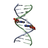
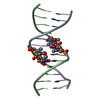
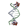
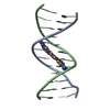
| Title | MOLECULAR STRUCTURE OF NICKED DNA. MODEL T4 | ||||||
 Components Components |
| ||||||
 Keywords Keywords |  DNA / B-DNA / DOUBLE HELIX / NICKED DNA / B-DNA / DOUBLE HELIX / NICKED | ||||||
| Function / homology | DNA / DNA (> 10) Function and homology information Function and homology information | ||||||
| Method | X-RAY DIFFRACTION / MOLECULAR REPLACEMENT / Resolution: 3 Å | ||||||
 Authors Authors | Aymani, J. / Coll, M. / Van Der Marel, G.A. / Van Boom, J.H. / Wang, A.H.-J. / Rich, A. | ||||||
 Citation Citation | Journal: Proc.Natl.Acad.Sci.USA / Year: 1990 Title: Molecular structure of nicked DNA: a substrate for DNA repair enzymes. Authors: Aymami, J. / Coll, M. / van der Marel, G.A. / van Boom, J.H. / Wang, A.H. / Rich, A. #1: Journal: Biochemistry / Year: 1989Title: Molecular Structure of the Netropsin-d(CGCGATATCGCG) Complex. DNA Conformation in an Alternating AT Segment Authors: Coll, M. / Aymami, J. / Van Der Marel, G.A. / Van Boom, J.H. / Rich, A. / Wang, A.H.-J. #2: Journal: Proc.Natl.Acad.Sci.USA / Year: 1987Title: A Bifurcated Hydrogen-Bonded Conformation in the d(*A(dot)*T) Base Pairs of the DNA Dodecamer d(CGCAAATTTGCG) and Its Complex With Distamycin Authors: Coll, M. / Frederick, C.A. / Wang, A.H.-J. / Rich, A. #3: Journal: Nature / Year: 1980Title: Crystal Structure Analysis of a Complete Turn of B-DNA Authors: Wing, R. / Drew, H. / Takano, T. / Broka, C. / Tanaka, S. / Itakura, K. / Dickerson, R.E. | ||||||
| History |
|
- Structure visualization
| Structure viewer | Molecule: MolmilJmol/JSmol |
|---|
- Downloads & links
-Download
| PDBx/mmCIF format | 1ndn.cif.gz | 23.3 KB | Display | PDBx/mmCIF format |
|---|---|---|---|---|
| PDB format | pdb1ndn.ent.gz | 13.7 KB | Display | PDB format |
| PDBx/mmJSON format | 1ndn.json.gz | Tree view | PDBx/mmJSON format | |
| Others |  Other downloads Other downloads |
-Validation report
| Arichive directory | https://data.pdbj.org/pub/pdb/validation_reports/nd/1ndnftp://data.pdbj.org/pub/pdb/validation_reports/nd/1ndn | HTTPS FTP |
|---|
-Related structure data


-Links
 PDBj
PDBj


- Assembly
| Deposited unit | 
| ||||||||
|---|---|---|---|---|---|---|---|---|---|
| 1 |
| ||||||||
| Unit cell |
|
-Components
| #1: DNA chain | Mass: 3681.420 Da / Num. of mol.: 1 / Source method: obtained synthetically |
|---|---|
| #2: DNA chain | Mass: 1800.203 Da / Num. of mol.: 1 / Source method: obtained synthetically |
| #3: DNA chain | Mass: 1800.203 Da / Num. of mol.: 1 / Source method: obtained synthetically |
| #4: Water | ChemComp-HOH / Water Mass: 18.015 Da / Num. of mol.: 61 / Source method: isolated from a natural source / Formula: H2O Mass: 18.015 Da / Num. of mol.: 61 / Source method: isolated from a natural source / Formula: H2O |
-Experimental details
-Experiment
| Experiment | Method: X-RAY DIFFRACTION |
|---|
- Sample preparation
| Crystal | Density Matthews: 2.62 Å3/Da / Density % sol: 53.01 % | ||||||||||||||||||||||||||||||||||||||||||||||||
|---|---|---|---|---|---|---|---|---|---|---|---|---|---|---|---|---|---|---|---|---|---|---|---|---|---|---|---|---|---|---|---|---|---|---|---|---|---|---|---|---|---|---|---|---|---|---|---|---|---|
| Crystal grow | Temperature: 277 K / Method: vapor diffusion / pH: 5 / Details: pH 5.00, VAPOR DIFFUSION, temperature 277.00K | ||||||||||||||||||||||||||||||||||||||||||||||||
| Components of the solutions |
| ||||||||||||||||||||||||||||||||||||||||||||||||
| Crystal grow | *PLUS Temperature: 4 ℃ / pH: 5 / Method: unknown | ||||||||||||||||||||||||||||||||||||||||||||||||
| Components of the solutions | *PLUS
|
-Data collection
| Diffraction | Mean temperature: 298 K |
|---|---|
| Diffraction source | Source: ROTATING ANODE |
| Detector | Type: RIGAKU AFC-5R / Detector: DIFFRACTOMETER |
| Radiation | Scattering type: x-ray |
| Radiation wavelength | Relative weight: 1 |
| Reflection | Highest resolution: 3 Å / Num. obs: 553 / Observed criterion σ(F): 2 |
| Reflection | *PLUS Highest resolution: 3 Å / Observed criterion σ(F): 2 |
- Processing
| Software |
| ||||||||||||
|---|---|---|---|---|---|---|---|---|---|---|---|---|---|
| Refinement | Method to determine structure: MOLECULAR REPLACEMENT / Highest resolution: 3 Å / σ(F): 2 /
| ||||||||||||
| Refine Biso |
| ||||||||||||
| Refinement step | Cycle: LAST / Highest resolution: 3 Å
|
