 Movie
Movie Controller

[English] 日本語
 Yorodumi
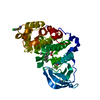
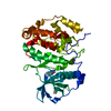
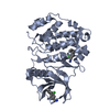
- PDB-1na7: Crystal structure of the catalytic subunit of human protein kinase CK2 -
+ Open data

- Basic information
| Entry | Database: PDB / ID: 1na7 | ||||||
|---|---|---|---|---|---|---|---|
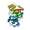
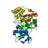
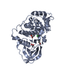
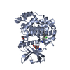
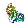
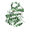
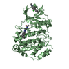
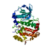
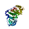
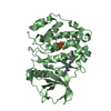
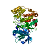
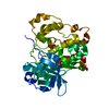
| Title | Crystal structure of the catalytic subunit of human protein kinase CK2 | ||||||
 Components Components | Casein kinase II, alpha chain | ||||||
 Keywords Keywords |  TRANSFERASE / CK2 / protein kinase / microcrystals / thin-film nanotechnology / microfocus ESRF TRANSFERASE / CK2 / protein kinase / microcrystals / thin-film nanotechnology / microfocus ESRF | ||||||
| Function / homology |  Function and homology information Function and homology informationregulation of chromosome separation / positive regulation of aggrephagy / WNT mediated activation of DVL / Condensation of Prometaphase Chromosomes / protein kinase CK2 complex / symbiont-mediated disruption of host cell PML body / Maturation of hRSV A proteins / Receptor Mediated Mitophagy / Sin3-type complex / Synthesis of PC ...regulation of chromosome separation / positive regulation of aggrephagy / WNT mediated activation of DVL / Condensation of Prometaphase Chromosomes / protein kinase CK2 complex / symbiont-mediated disruption of host cell PML body / Maturation of hRSV A proteins / Receptor Mediated Mitophagy / Sin3-type complex / Synthesis of PC / RUNX1 interacts with co-factors whose precise effect on RUNX1 targets is not known / negative regulation of apoptotic signaling pathway / positive regulation of Wnt signaling pathway / negative regulation of double-strand break repair via homologous recombination / chaperone-mediated protein folding / negative regulation of ubiquitin-dependent protein catabolic process / Signal transduction by L1 / peptidyl-threonine phosphorylation / Hsp90 protein binding / negative regulation of cysteine-type endopeptidase activity involved in apoptotic process / PML body / Wnt signaling pathway / Regulation of PTEN stability and activity / Cooperation of PDCL (PhLP1) and TRiC/CCT in G-protein beta folding / positive regulation of protein catabolic process / KEAP1-NFE2L2 pathway / double-strand break repair / rhythmic process / kinase activity / positive regulation of cell growth / peptidyl-serine phosphorylation / Regulation of TP53 Activity through Phosphorylation / negative regulation of translation / protein stabilization / non-specific serine/threonine protein kinase / regulation of cell cycle / cell cycle / protein phosphorylation / protein serine kinase activity / protein serine/threonine kinase activity / apoptotic process / DNA damage response / positive regulation of cell population proliferation / signal transduction / nucleoplasm / ATP binding / identical protein binding / nucleus / plasma membrane / cytosolSimilarity search - Function | ||||||
| Biological species |  Homo sapiens (human) Homo sapiens (human) | ||||||
| Method | X-RAY DIFFRACTION / SYNCHROTRON / MOLECULAR REPLACEMENT / Resolution: 2.4 Å | ||||||
 Authors Authors | Pechkova, E. / Zanotti, G. / Nicolini, C. | ||||||
 Citation Citation | Journal: Acta Crystallogr.,Sect.D / Year: 2003 Title: Three-dimensional atomic structure of a catalytic subunit mutant of human protein kinase CK2. Authors: Pechkova, E. / Zanotti, G. / Nicolini, C. #1: Journal: Embo J. / Year: 1998Title: Crystal structure of the catalytic subunit of protein kinase CK2 from Zea mays at 2.1 A resolution Authors: Niefind, K. / Guerra, B. / Pinna, L.A. / Issinger, O.-G. / Schomburg, D. #2: Journal: Protein Sci. / Year: 2001Title: Structural features underlying selective inhibition of protein kinase CK2 by ATP site-directed tetrabromo-2-benzotriazole Authors: Battistutta, R. / De Moliner, E. / Sarno, S. / Zanotti, G. / Pinna, L.A. #3: Journal: J.CRYST.GROWTH / Year: 2001Title: Accelerated protein crystal growth by protein thin film template Authors: Pechkova, E. / Nicolini, C. #4: Journal: J.Cell.Biochem. / Year: 2002Title: Protein nucleation and crystallization by homologous protein thin film template Authors: Pechkova, E. / Nicolini, C. #5: Journal: Nanotechnology / Year: 2002Title: From art to science in protein crystallization by means of thin-film technology Authors: Pechkova, E. / Nicolini, C. | ||||||
| History |
|
- Structure visualization
| Structure viewer | Molecule: MolmilJmol/JSmol |
|---|
- Downloads & links
-Download
| PDBx/mmCIF format | 1na7.cif.gz | 83.1 KB | Display | PDBx/mmCIF format |
|---|---|---|---|---|
| PDB format | pdb1na7.ent.gz | 62.1 KB | Display | PDB format |
| PDBx/mmJSON format | 1na7.json.gz | Tree view | PDBx/mmJSON format | |
| Others |  Other downloads Other downloads |
-Validation report
| Arichive directory | https://data.pdbj.org/pub/pdb/validation_reports/na/1na7ftp://data.pdbj.org/pub/pdb/validation_reports/na/1na7 | HTTPS FTP |
|---|
-Related structure data
| Related structure data |  1m2rS S: Starting model for refinement |
|---|---|
| Similar structure data |


-Links
 PDBj
PDBj


- Assembly
| Deposited unit | 
| ||||||||
|---|---|---|---|---|---|---|---|---|---|
| 1 |
| ||||||||
| Unit cell |
|
-Components
| #1: Protein | Mass: 39333.895 Da / Num. of mol.: 1 / Fragment: Catalytic subunit / Mutation: E27A, K76N Source method: isolated from a genetically manipulated source Source: (gene. exp.) Homo sapiens (human) / Species (production host): Escherichia coli / Production host:  Escherichia coli BL21 (bacteria) / Strain (production host): BL21 / References: UniProt: P68400, EC: 2.7.1.37 Escherichia coli BL21 (bacteria) / Strain (production host): BL21 / References: UniProt: P68400, EC: 2.7.1.37 |
|---|---|
| #2: Water | ChemComp-HOH / Water Mass: 18.015 Da / Num. of mol.: 91 / Source method: isolated from a natural source / Formula: H2O Mass: 18.015 Da / Num. of mol.: 91 / Source method: isolated from a natural source / Formula: H2O |
-Experimental details
-Experiment
| Experiment | Method: X-RAY DIFFRACTION / Number of used crystals: 1 |
|---|
- Sample preparation
| Crystal | Density Matthews: 2.03 Å3/Da / Density % sol: 39.54 % | ||||||||||||||||||||||||||||||
|---|---|---|---|---|---|---|---|---|---|---|---|---|---|---|---|---|---|---|---|---|---|---|---|---|---|---|---|---|---|---|---|
| Crystal grow | Temperature: 293 K / Method: lb modified hanging drop method / pH: 8 Details: PEG 3500, NaAC, Tris, pH 8, LB Modified hanging drop method, temperature 293K | ||||||||||||||||||||||||||||||
| Crystal grow | *PLUS Temperature: 293 K / Method: vapor diffusion, hanging drop | ||||||||||||||||||||||||||||||
| Components of the solutions | *PLUS
|
-Data collection
| Diffraction | Mean temperature: 100 K |
|---|---|
| Diffraction source | Source: SYNCHROTRON / Site: ESRF  / Beamline: ID13 / Wavelength: 0.9755 Å / Beamline: ID13 / Wavelength: 0.9755 Å |
| Detector | Type: MARRESEARCH / Detector: CCD / Date: Nov 10, 2002 / Details: microfocus |
| Radiation | Protocol: SINGLE WAVELENGTH / Monochromatic (M) / Laue (L): M / Scattering type: x-ray |
| Radiation wavelength | Wavelength: 0.9755 Å / Relative weight: 1 |
| Reflection | Resolution: 2.4→29.21 Å / Num. all: 12457 / Num. obs: 12457 / % possible obs: 99 % / Observed criterion σ(F): 0 / Observed criterion σ(I): 0 / Redundancy: 2.3 % / Biso Wilson estimate: 21 Å2 / Rmerge(I) obs: 0.13 / Net I/σ(I): 4.3 |
| Reflection shell | Resolution: 2.4→2.53 Å / Redundancy: 2.3 % / Rmerge(I) obs: 0.37 / Mean I/σ(I) obs: 1.6 / Num. unique all: 1817 / % possible all: 99.4 |
| Reflection | *PLUS Highest resolution: 2.4 Å / Lowest resolution: 29 Å / % possible obs: 99 % |
| Reflection shell | *PLUS % possible obs: 99.4 % / Num. unique obs: 1817 |
- Processing
| Software |
| ||||||||||||||||||||||||||||||||||||||||||||||||||||||||||||
|---|---|---|---|---|---|---|---|---|---|---|---|---|---|---|---|---|---|---|---|---|---|---|---|---|---|---|---|---|---|---|---|---|---|---|---|---|---|---|---|---|---|---|---|---|---|---|---|---|---|---|---|---|---|---|---|---|---|---|---|---|---|
| Refinement | Method to determine structure: MOLECULAR REPLACEMENT Starting model: PDB ENTRY 1M2R Resolution: 2.4→29.21 Å / Rfactor Rfree error: 0.008 / Isotropic thermal model: RESTRAINED / Cross valid method: THROUGHOUT / σ(F): 0 / Stereochemistry target values: Engh & Huber
| ||||||||||||||||||||||||||||||||||||||||||||||||||||||||||||
| Solvent computation | Solvent model: FLAT MODEL / Bsol: 31.8231 Å2 / ksol: 0.360107 e/Å3 | ||||||||||||||||||||||||||||||||||||||||||||||||||||||||||||
| Displacement parameters | Biso mean: 27.6 Å2
| ||||||||||||||||||||||||||||||||||||||||||||||||||||||||||||
| Refine analyze | Luzzati coordinate error free: 0.38 Å / Luzzati sigma a free: 0.31 Å | ||||||||||||||||||||||||||||||||||||||||||||||||||||||||||||
| Refinement step | Cycle: LAST / Resolution: 2.4→29.21 Å
| ||||||||||||||||||||||||||||||||||||||||||||||||||||||||||||
| Refine LS restraints |
| ||||||||||||||||||||||||||||||||||||||||||||||||||||||||||||
| LS refinement shell | Resolution: 2.4→2.55 Å / Rfactor Rfree error: 0.02 / Total num. of bins used: 6
| ||||||||||||||||||||||||||||||||||||||||||||||||||||||||||||
| Xplor file |
| ||||||||||||||||||||||||||||||||||||||||||||||||||||||||||||
| Refinement | *PLUS Lowest resolution: 29 Å / Num. reflection obs: 12457 | ||||||||||||||||||||||||||||||||||||||||||||||||||||||||||||
| Solvent computation | *PLUS | ||||||||||||||||||||||||||||||||||||||||||||||||||||||||||||
| Displacement parameters | *PLUS | ||||||||||||||||||||||||||||||||||||||||||||||||||||||||||||
| Refine LS restraints | *PLUS
| ||||||||||||||||||||||||||||||||||||||||||||||||||||||||||||
| LS refinement shell | *PLUS Lowest resolution: 2.53 Å / Rfactor Rfree: 0.03 / Num. reflection Rwork: 1817 |
