 Movie
Movie Controller


+ Open data

- Basic information
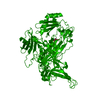
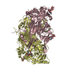
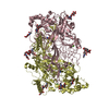
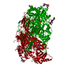
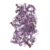
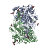
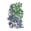
| Entry | Database: PDB / ID: 1n9e | |||||||||
|---|---|---|---|---|---|---|---|---|---|---|
| Title | Crystal structure of Pichia pastoris Lysyl Oxidase PPLO | |||||||||
 Components Components | LYSYL OXIDASE | |||||||||
 Keywords Keywords | OXIDOREDUCTASE / AMINE OXIDASE / QUINOPROTEIN / TOPAQUINONE ENZYME / TPQ | |||||||||
| Function / homology |  Function and homology informationOxidoreductases; Acting on the CH-NH2 group of donors; With oxygen as acceptor / diamine oxidase activity / primary amine oxidase activity / amine metabolic process / quinone binding / copper ion binding / plasma membrane Function and homology informationOxidoreductases; Acting on the CH-NH2 group of donors; With oxygen as acceptor / diamine oxidase activity / primary amine oxidase activity / amine metabolic process / quinone binding / copper ion binding / plasma membraneSimilarity search - Function | |||||||||
| Biological species |  Pichia pastoris (fungus) Pichia pastoris (fungus) | |||||||||
| Method | X-RAY DIFFRACTION / SYNCHROTRON / MOLECULAR REPLACEMENT / Resolution: 1.65 Å | |||||||||
 Authors Authors | Guss, J.M. / Duff, A.P. | |||||||||
 Citation Citation | Journal: Biochemistry / Year: 2003 Title: The Crystal Structure of Pichia pastoris Lysyl Oxidase Authors: Duff, A.P. / Cohen, A.E. / Ellis, P.J. / Kuchar, J.A. / Langley, D.B. / Shepard, E.M. / Dooley, D.M. / Freeman, H.C. / Guss, J.M. | |||||||||
| History |
|
- Structure visualization
| Structure viewer | Molecule: MolmilJmol/JSmol |
|---|
- Downloads & links
-Download
| PDBx/mmCIF format | 1n9e.cif.gz | 695.6 KB | Display | PDBx/mmCIF format |
|---|---|---|---|---|
| PDB format | pdb1n9e.ent.gz | 565.5 KB | Display | PDB format |
| PDBx/mmJSON format | 1n9e.json.gz | Tree view | PDBx/mmJSON format | |
| Others |  Other downloads Other downloads |
-Validation report
| Arichive directory | https://data.pdbj.org/pub/pdb/validation_reports/n9/1n9eftp://data.pdbj.org/pub/pdb/validation_reports/n9/1n9e | HTTPS FTP |
|---|
-Related structure data
| Related structure data |  1oacS S: Starting model for refinement |
|---|---|
| Similar structure data |


-Links
 PDBj
PDBj- Assembly
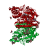
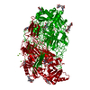
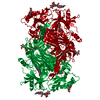
| Deposited unit | 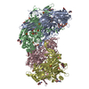
| ||||||||
|---|---|---|---|---|---|---|---|---|---|
| 1 | 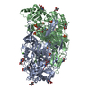
| ||||||||
| 2 | 
| ||||||||
| Unit cell |
|
-Components
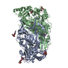
-Protein , 1 types, 4 molecules ABCD
| #1: Protein | Mass: 89800.625 Da / Num. of mol.: 4 / Source method: isolated from a natural source / Source: (natural) Pichia pastoris (fungus)References: GenBank: 13936870, UniProt: Q96X16*PLUS, protein-lysine 6-oxidase |
|---|
-Sugars , 2 types, 20 molecules 
| #2: Polysaccharide | 2-acetamido-2-deoxy-beta-D-glucopyranose-(1-4)-2-acetamido-2-deoxy-beta-D-glucopyranose / Mass: 424.401 Da / Num. of mol.: 4Source method: isolated from a genetically manipulated source #3: Sugar | ChemComp-NAG / N-Acetylglucosamine Type: D-saccharide, beta linking / Mass: 221.208 Da / Num. of mol.: 16 Type: D-saccharide, beta linking / Mass: 221.208 Da / Num. of mol.: 16Source method: isolated from a genetically manipulated source Formula: C8H15NO6 |
|---|
-Non-polymers , 4 types, 4021 molecules 


| #4: Chemical | ChemComp-CU / Copper Mass: 63.546 Da / Num. of mol.: 4 / Source method: obtained synthetically / Formula: Cu Mass: 63.546 Da / Num. of mol.: 4 / Source method: obtained synthetically / Formula: Cu#5: Chemical | ChemComp-CA /  Mass: 40.078 Da / Num. of mol.: 8 / Source method: obtained synthetically / Formula: Ca Mass: 40.078 Da / Num. of mol.: 8 / Source method: obtained synthetically / Formula: Ca#6: Chemical | ChemComp-SO4 / Sulfate Mass: 96.063 Da / Num. of mol.: 28 / Source method: obtained synthetically / Formula: SO4 Mass: 96.063 Da / Num. of mol.: 28 / Source method: obtained synthetically / Formula: SO4#7: Water | ChemComp-HOH / | WaterMass: 18.015 Da / Num. of mol.: 3981 / Source method: isolated from a natural source / Formula: H2O |
|---|
-Experimental details
-Experiment
| Experiment | Method: X-RAY DIFFRACTION / Number of used crystals: 1 |
|---|
- Sample preparation
| Crystal | Density Matthews: 2.34 Å3/Da / Density % sol: 47 % | ||||||||||||||||||||
|---|---|---|---|---|---|---|---|---|---|---|---|---|---|---|---|---|---|---|---|---|---|
| Crystal grow | Temperature: 298 K / Method: vapor diffusion / pH: 7 Details: 25% PEG 4000, 0.175M ammonium sulfate, 15% MPD, VAPOR DIFFUSION, temperature 298K | ||||||||||||||||||||
| Crystal grow | *PLUS Temperature: 277 K / Method: vapor diffusion, hanging drop / Details: Lee, M., (2002) Acta Cryst., D58, 2177. | ||||||||||||||||||||
| Components of the solutions | *PLUS
|
-Data collection
| Diffraction | Mean temperature: 100 K |
|---|---|
| Diffraction source | Source: SYNCHROTRON / Site: SSRL  / Beamline: BL9-1 / Wavelength: 0.99184 / Beamline: BL9-1 / Wavelength: 0.99184 |
| Detector | Type: MAR scanner 345 mm plate / Detector: IMAGE PLATE / Date: Dec 17, 2001 |
| Radiation | Protocol: SINGLE WAVELENGTH / Monochromatic (M) / Laue (L): M / Scattering type: x-ray |
| Radiation wavelength | Wavelength: 0.99184 Å / Relative weight: 1 |
| Reflection | Resolution: 1.65→24.6 Å / Num. obs: 419014 / % possible obs: 94.7 % / Redundancy: 2.8 % / Rmerge(I) obs: 0.062 / Net I/σ(I): 9.1 |
| Reflection shell | Resolution: 1.65→1.69 Å / Redundancy: 2.6 % / Rmerge(I) obs: 0.42 / Mean I/σ(I) obs: 2.2 |
| Reflection | *PLUS Highest resolution: 1.65 Å / Lowest resolution: 24.6 Å / Redundancy: 2.8 % / Rmerge(I) obs: 0.06 |
| Reflection shell | *PLUS Lowest resolution: 1.68 Å / % possible obs: 90.7 % / Redundancy: 2.6 % / Num. unique obs: 28231 / Mean I/σ(I) obs: 2.4 |
- Processing
| Software |
| ||||||||||||||||||||||||||||||||||||||||||||||||||||||||||||||||||||||||||||||||||||||||||||||||||||||||||||||||||||||||||||||||||||||||||||||||||||||||||||||||
|---|---|---|---|---|---|---|---|---|---|---|---|---|---|---|---|---|---|---|---|---|---|---|---|---|---|---|---|---|---|---|---|---|---|---|---|---|---|---|---|---|---|---|---|---|---|---|---|---|---|---|---|---|---|---|---|---|---|---|---|---|---|---|---|---|---|---|---|---|---|---|---|---|---|---|---|---|---|---|---|---|---|---|---|---|---|---|---|---|---|---|---|---|---|---|---|---|---|---|---|---|---|---|---|---|---|---|---|---|---|---|---|---|---|---|---|---|---|---|---|---|---|---|---|---|---|---|---|---|---|---|---|---|---|---|---|---|---|---|---|---|---|---|---|---|---|---|---|---|---|---|---|---|---|---|---|---|---|---|---|---|---|
| Refinement | Method to determine structure: MOLECULAR REPLACEMENT Starting model: PDB ENTRY 1OAC Resolution: 1.65→24.6 Å / Cor.coef. Fo:Fc: 0.964 / SU B: 1.493 / SU ML: 0.05 / Cross valid method: THROUGHOUT, EXCEPT FINAL ROUND / ESU R: 0.082 / Stereochemistry target values: MAXIMUM LIKELIHOOD Details: THE STRUCTURE WAS ALSO REFINED WITH CNS VER.1.1 SO4 811, 821, 831 AND 841 ARE MODELLED WITH ZERO OCCUPANCY. ELECTRON DENSITY FOR THESE IONS STRONGLY SUGGESTS THEIR PRESENCE, HOWEVER, THE ...Details: THE STRUCTURE WAS ALSO REFINED WITH CNS VER.1.1 SO4 811, 821, 831 AND 841 ARE MODELLED WITH ZERO OCCUPANCY. ELECTRON DENSITY FOR THESE IONS STRONGLY SUGGESTS THEIR PRESENCE, HOWEVER, THE MODELLED POSITIONS ARE NOT STABLE IN POSITIONAL REFINEMENT, AND RESULTING REFINED POSITIONS HAVE UNACCEPTABLE VAN DER WAALS VIOLATIONS WITH NEIGHBOURING PROTEIN ATOMS. FREE_R WAS MONITORED IN ALL ROUNDS OF REFINEMENT EXCEPT THE LAST. THE TEST SET SIZE WAS 5%, AND WAS SELECTED RANDOMLY. IN THE LAST ROUND OF REFINEMENT, THE MODEL WAS REFINED AGAINST ALL OF THE DATA TO PRODUCE THE FINAL MODEL.
| ||||||||||||||||||||||||||||||||||||||||||||||||||||||||||||||||||||||||||||||||||||||||||||||||||||||||||||||||||||||||||||||||||||||||||||||||||||||||||||||||
| Solvent computation | Ion probe radii: 0.8 Å / Shrinkage radii: 0.8 Å / VDW probe radii: 1.4 Å / Solvent model: BABINET MODEL WITH MASK | ||||||||||||||||||||||||||||||||||||||||||||||||||||||||||||||||||||||||||||||||||||||||||||||||||||||||||||||||||||||||||||||||||||||||||||||||||||||||||||||||
| Displacement parameters | Biso mean: 17.86 Å2
| ||||||||||||||||||||||||||||||||||||||||||||||||||||||||||||||||||||||||||||||||||||||||||||||||||||||||||||||||||||||||||||||||||||||||||||||||||||||||||||||||
| Refinement step | Cycle: LAST / Resolution: 1.65→24.6 Å
| ||||||||||||||||||||||||||||||||||||||||||||||||||||||||||||||||||||||||||||||||||||||||||||||||||||||||||||||||||||||||||||||||||||||||||||||||||||||||||||||||
| Refine LS restraints |
| ||||||||||||||||||||||||||||||||||||||||||||||||||||||||||||||||||||||||||||||||||||||||||||||||||||||||||||||||||||||||||||||||||||||||||||||||||||||||||||||||
| LS refinement shell | Resolution: 1.65→1.69 Å / Total num. of bins used: 20 /
| ||||||||||||||||||||||||||||||||||||||||||||||||||||||||||||||||||||||||||||||||||||||||||||||||||||||||||||||||||||||||||||||||||||||||||||||||||||||||||||||||
| Software | *PLUS Version: 5 / Classification: refinement | ||||||||||||||||||||||||||||||||||||||||||||||||||||||||||||||||||||||||||||||||||||||||||||||||||||||||||||||||||||||||||||||||||||||||||||||||||||||||||||||||
| Refinement | *PLUS Lowest resolution: 24.6 Å / % reflection Rfree: 5 % / Rfactor all: 0.16 / Rfactor Rfree: 0.187 / Rfactor Rwork: 0.161 | ||||||||||||||||||||||||||||||||||||||||||||||||||||||||||||||||||||||||||||||||||||||||||||||||||||||||||||||||||||||||||||||||||||||||||||||||||||||||||||||||
| Solvent computation | *PLUS | ||||||||||||||||||||||||||||||||||||||||||||||||||||||||||||||||||||||||||||||||||||||||||||||||||||||||||||||||||||||||||||||||||||||||||||||||||||||||||||||||
| Displacement parameters | *PLUS | ||||||||||||||||||||||||||||||||||||||||||||||||||||||||||||||||||||||||||||||||||||||||||||||||||||||||||||||||||||||||||||||||||||||||||||||||||||||||||||||||
| Refine LS restraints | *PLUS
| ||||||||||||||||||||||||||||||||||||||||||||||||||||||||||||||||||||||||||||||||||||||||||||||||||||||||||||||||||||||||||||||||||||||||||||||||||||||||||||||||
| LS refinement shell | *PLUS Lowest resolution: 1.68 Å / Rfactor Rwork: 0.24 / Rfactor obs: 0.23 |
