 Movie
Movie Controller

[English] 日本語
 Yorodumi
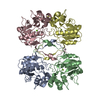
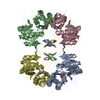
- PDB-1n8n: Crystal structure of the Au3+ complex of AphA class B acid phosph... -
+ Open data

- Basic information
| Entry | Database: PDB / ID: 1n8n | ||||||
|---|---|---|---|---|---|---|---|
| Title | Crystal structure of the Au3+ complex of AphA class B acid phosphatase/phosphotransferase from E. coli at 1.69 A resolution | ||||||
 Components Components | Class B acid phosphatase | ||||||
 Keywords Keywords |  HYDROLASE / Class B acid phosphatase / DDDD acid phosphatase / metallo-enzyme HYDROLASE / Class B acid phosphatase / DDDD acid phosphatase / metallo-enzyme | ||||||
| Function / homology |  Function and homology information Function and homology informationL-phosphoserine phosphatase activity / acid phosphatase / acid phosphatase activity / outer membrane-bounded periplasmic space / magnesium ion binding / identical protein binding / metal ion bindingSimilarity search - Function | ||||||
| Biological species |  Escherichia coli (E. coli) Escherichia coli (E. coli) | ||||||
| Method | X-RAY DIFFRACTION / SYNCHROTRON / MOLECULAR REPLACEMENT / Resolution: 1.69 Å | ||||||
 Authors Authors | Calderone, V. / Forleo, C. / Benvenuti, M. / Rossolini, G.M. / Thaller, M.C. / Mangani, S. | ||||||
 Citation Citation | Journal: J.Mol.Biol. / Year: 2004 Title: The first structure of a bacterial class B Acid phosphatase reveals further structural heterogeneity among phosphatases of the haloacid dehalogenase fold. Authors: Calderone, V. / Forleo, C. / Benvenuti, M. / Thaller, M.C. / Rossolini, G.M. / Mangani, S. #1: Journal: FEMS Microbiol.Lett. / Year: 1997Title: Identification of the gene (aphA) encoding the class B acid phosphatase/phosphotransferase of Escherichia coli MG1655 and characterization of its product Authors: Thaller, M.C. / Schippa, S. / Bonci, A. / Cresti, S. / Rossolini, G.M. | ||||||
| History |
|
- Structure visualization
| Structure viewer | Molecule: MolmilJmol/JSmol |
|---|
- Downloads & links
-Download
| PDBx/mmCIF format | 1n8n.cif.gz | 61.6 KB | Display | PDBx/mmCIF format |
|---|---|---|---|---|
| PDB format | pdb1n8n.ent.gz | 44.8 KB | Display | PDB format |
| PDBx/mmJSON format | 1n8n.json.gz | Tree view | PDBx/mmJSON format | |
| Others |  Other downloads Other downloads |
-Validation report
| Arichive directory | https://data.pdbj.org/pub/pdb/validation_reports/n8/1n8nftp://data.pdbj.org/pub/pdb/validation_reports/n8/1n8n | HTTPS FTP |
|---|
-Related structure data


-Links
 PDBj
PDBj
- Assembly
| Deposited unit | 
| ||||||||
|---|---|---|---|---|---|---|---|---|---|
| 1 | 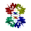
| ||||||||
| Unit cell |
| ||||||||
| Components on special symmetry positions |
| ||||||||
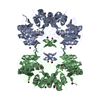
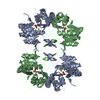
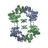
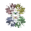
| Details | The biological assembly is a tetramer |
-Components
| #1: Protein | Mass: 23555.342 Da / Num. of mol.: 1 Source method: isolated from a genetically manipulated source Source: (gene. exp.) Escherichia coli (E. coli) / Gene: APHA / Production host: Escherichia coli (E. coli)References: UniProt: P32697, UniProt: P0AE22*PLUS, acid phosphatase |
|---|---|
| #2: Chemical | ChemComp-AU3 /   Mass: 196.967 Da / Num. of mol.: 1 / Source method: obtained synthetically / Formula: Au Mass: 196.967 Da / Num. of mol.: 1 / Source method: obtained synthetically / Formula: Au |
| #3: Water | ChemComp-HOH / Water Mass: 18.015 Da / Num. of mol.: 284 / Source method: isolated from a natural source / Formula: H2O Mass: 18.015 Da / Num. of mol.: 284 / Source method: isolated from a natural source / Formula: H2O |
-Experimental details
-Experiment
| Experiment | Method: X-RAY DIFFRACTION / Number of used crystals: 1 |
|---|
- Sample preparation
| Crystal | Density Matthews: 3.34 Å3/Da / Density % sol: 63.16 % | ||||||||||||||||||||||||
|---|---|---|---|---|---|---|---|---|---|---|---|---|---|---|---|---|---|---|---|---|---|---|---|---|---|
| Crystal grow | Temperature: 293 K / Method: vapor diffusion, sitting drop / pH: 7.2 Details: AphA 6mg/mL, 50 mM Na acetate, 25% PEG 6000, pH 7.2, VAPOR DIFFUSION, SITTING DROP, temperature 293K. The crystals of the native enzyme (containing Mg2+ or Zn2+) have been soaked in 1 mM solution of AuCl3 | ||||||||||||||||||||||||
| Crystal grow | *PLUS Method: vapor diffusion, sitting drop | ||||||||||||||||||||||||
| Components of the solutions | *PLUS
|
-Data collection
| Diffraction | Mean temperature: 100 K | ||||||||||||
|---|---|---|---|---|---|---|---|---|---|---|---|---|---|
| Diffraction source | Source: SYNCHROTRON / Site: ESRF  / Beamline: ID14-4 / Wavelength: 1.0377, 1.0404, 0.9392 / Beamline: ID14-4 / Wavelength: 1.0377, 1.0404, 0.9392 | ||||||||||||
| Detector | Type: ADSC QUANTUM 4 / Detector: CCD / Date: Jul 13, 2002 / Details: Toroidal mirror | ||||||||||||
| Radiation | Monochromator: Double crystal, Si(111) / Protocol: MAD / Monochromatic (M) / Laue (L): M / Scattering type: x-ray | ||||||||||||
| Radiation wavelength |
| ||||||||||||
| Reflection | Resolution: 1.69→46.13 Å / Num. all: 31884 / Num. obs: 31884 / % possible obs: 88.8 % / Observed criterion σ(F): 0 / Observed criterion σ(I): 0 / Redundancy: 3.7 % / Biso Wilson estimate: 19.709 Å2 / Rmerge(I) obs: 0.061 / Rsym value: 0.061 / Net I/σ(I): 7 | ||||||||||||
| Reflection shell | Resolution: 1.69→1.79 Å / Redundancy: 1.6 % / Rmerge(I) obs: 0.093 / Mean I/σ(I) obs: 7 / Num. unique all: 2976 / Rsym value: 0.093 / % possible all: 53 | ||||||||||||
| Reflection | *PLUS Lowest resolution: 25 Å / % possible obs: 98.2 % / Redundancy: 4.3 % / Num. measured all: 135549 / Rmerge(I) obs: 0.075 | ||||||||||||
| Reflection shell | *PLUS % possible obs: 92 % / Redundancy: 2.9 % / Num. unique obs: 2579 / Num. measured obs: 7369 / Rmerge(I) obs: 0.375 / Mean I/σ(I) obs: 1.1 |
- Processing
| Software |
| ||||||||||||||||||||||||||||||||||||||||||||||||||||||||||||||||||||||
|---|---|---|---|---|---|---|---|---|---|---|---|---|---|---|---|---|---|---|---|---|---|---|---|---|---|---|---|---|---|---|---|---|---|---|---|---|---|---|---|---|---|---|---|---|---|---|---|---|---|---|---|---|---|---|---|---|---|---|---|---|---|---|---|---|---|---|---|---|---|---|---|
| Refinement | Method to determine structure: MOLECULAR REPLACEMENT Starting model: MAD-phased Bromine derivative at 2.2 A resolution Resolution: 1.69→46.13 Å / Cor.coef. Fo:Fc: 0.956 / Cor.coef. Fo:Fc free: 0.941 / Cross valid method: THROUGHOUT / σ(F): 0 / σ(I): 0 / Stereochemistry target values: MAXIMUM LIKELIHOOD
| ||||||||||||||||||||||||||||||||||||||||||||||||||||||||||||||||||||||
| Solvent computation | Shrinkage radii: 0.8 Å / Solvent model: BABINET MODEL WITH MASK | ||||||||||||||||||||||||||||||||||||||||||||||||||||||||||||||||||||||
| Displacement parameters | Biso mean: 20.602 Å2
| ||||||||||||||||||||||||||||||||||||||||||||||||||||||||||||||||||||||
| Refine analyze | Luzzati sigma a obs: 0.23 Å | ||||||||||||||||||||||||||||||||||||||||||||||||||||||||||||||||||||||
| Refinement step | Cycle: LAST / Resolution: 1.69→46.13 Å
| ||||||||||||||||||||||||||||||||||||||||||||||||||||||||||||||||||||||
| Refine LS restraints |
| ||||||||||||||||||||||||||||||||||||||||||||||||||||||||||||||||||||||
| LS refinement shell | Resolution: 1.69→1.79 Å / Total num. of bins used: 20
| ||||||||||||||||||||||||||||||||||||||||||||||||||||||||||||||||||||||
| Refinement | *PLUS Lowest resolution: 25 Å / Rfactor Rfree: 0.207 / Rfactor Rwork: 0.178 | ||||||||||||||||||||||||||||||||||||||||||||||||||||||||||||||||||||||
| Solvent computation | *PLUS | ||||||||||||||||||||||||||||||||||||||||||||||||||||||||||||||||||||||
| Displacement parameters | *PLUS | ||||||||||||||||||||||||||||||||||||||||||||||||||||||||||||||||||||||
| Refine LS restraints | *PLUS
|
