 Movie
Movie Controller

[English] 日本語
 Yorodumi
- PDB-1n7j: The structure of Phenylethanolamine N-methyltransferase in comple... -
+ Open data

- Basic information
| Entry | Database: PDB / ID: 1n7j | ||||||
|---|---|---|---|---|---|---|---|
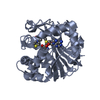
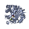
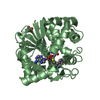
| Title | The structure of Phenylethanolamine N-methyltransferase in complex with S-adenosylhomocysteine and an iodinated inhibitor | ||||||
 Components Components | Phenylethanolamine N-methyltransferase | ||||||
 Keywords Keywords | TRANSFERASE / methyltransferase / catecholamine / adrenaline / epinephrine / s-adenosylmethionine / s-adenolsylhomocysteine | ||||||
| Function / homology |  Function and homology informationphenylethanolamine N-methyltransferase / phenylethanolamine N-methyltransferase activity / epinephrine biosynthetic process / Catecholamine biosynthesis / catecholamine biosynthetic process / methylation / cytosol Function and homology informationphenylethanolamine N-methyltransferase / phenylethanolamine N-methyltransferase activity / epinephrine biosynthetic process / Catecholamine biosynthesis / catecholamine biosynthetic process / methylation / cytosolSimilarity search - Function | ||||||
| Biological species |  Homo sapiens (human) Homo sapiens (human) | ||||||
| Method | X-RAY DIFFRACTION / difference fourier / Resolution: 2.7 Å | ||||||
 Authors Authors | McMillan, F.M. / Archbold, J. / McLeish, M.J. / Caine, J.M. / Criscione, K.R. / Grunewald, G.L. / Martin, J.L. | ||||||
 Citation Citation | Journal: J.Med.Chem. / Year: 2004 Title: Molecular recognition of sub-micromolar inhibitors by the epinephrine-synthesizing enzyme phenylethanolamine N-methyltransferase. Authors: McMillan, F.M. / Archbold, J. / McLeish, M.J. / Caine, J.M. / Criscione, K.R. / Grunewald, G.L. / Martin, J.L. | ||||||
| History |
|
- Structure visualization
| Structure viewer | Molecule: MolmilJmol/JSmol |
|---|
- Downloads & links
-Download
| PDBx/mmCIF format | 1n7j.cif.gz | 116.1 KB | Display | PDBx/mmCIF format |
|---|---|---|---|---|
| PDB format | pdb1n7j.ent.gz | 89.3 KB | Display | PDB format |
| PDBx/mmJSON format | 1n7j.json.gz | Tree view | PDBx/mmJSON format | |
| Others |  Other downloads Other downloads |
-Validation report
| Arichive directory | https://data.pdbj.org/pub/pdb/validation_reports/n7/1n7jftp://data.pdbj.org/pub/pdb/validation_reports/n7/1n7j | HTTPS FTP |
|---|
-Related structure data
| Related structure data | 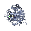 1n7iC 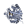 1hnnS S: Starting model for refinement C: citing same article ( |
|---|---|
| Similar structure data |


-Links
 PDBj
PDBj- Assembly
| Deposited unit | 
| ||||||||
|---|---|---|---|---|---|---|---|---|---|
| 1 | 
| ||||||||
| 2 | 
| ||||||||
| Unit cell |
|
-Components
| #1: Protein | / PNMTase / Noradrenaline N-methyltransferase Mass: 30887.982 Da / Num. of mol.: 2 Source method: isolated from a genetically manipulated source Source: (gene. exp.) Homo sapiens (human) / Gene: PNMT OR PENT / Plasmid: pET17 / Species (production host): Escherichia coli / Production host:  Escherichia coli BL21(DE3) (bacteria) / Strain (production host): BL21(DE3) Escherichia coli BL21(DE3) (bacteria) / Strain (production host): BL21(DE3)References: UniProt: P11086, phenylethanolamine N-methyltransferase#2: Chemical | S-Adenosyl-L-homocysteine  Type: L-peptide linking / Mass: 384.411 Da / Num. of mol.: 2 / Source method: obtained synthetically / Formula: C14H20N6O5S Type: L-peptide linking / Mass: 384.411 Da / Num. of mol.: 2 / Source method: obtained synthetically / Formula: C14H20N6O5S#3: Chemical |   Mass: 259.087 Da / Num. of mol.: 2 / Source method: obtained synthetically / Formula: C9H10IN Mass: 259.087 Da / Num. of mol.: 2 / Source method: obtained synthetically / Formula: C9H10IN#4: Water | ChemComp-HOH / | Water Mass: 18.015 Da / Num. of mol.: 56 / Source method: isolated from a natural source / Formula: H2O Mass: 18.015 Da / Num. of mol.: 56 / Source method: isolated from a natural source / Formula: H2O |
|---|
-Experimental details
-Experiment
| Experiment | Method: X-RAY DIFFRACTION / Number of used crystals: 1 |
|---|
- Sample preparation
| Crystal | Density Matthews: 3.53 Å3/Da / Density % sol: 64.86 % | ||||||||||||||||||||||||||||||||||||||||||||||||||||||||
|---|---|---|---|---|---|---|---|---|---|---|---|---|---|---|---|---|---|---|---|---|---|---|---|---|---|---|---|---|---|---|---|---|---|---|---|---|---|---|---|---|---|---|---|---|---|---|---|---|---|---|---|---|---|---|---|---|---|
| Crystal grow | Temperature: 293 K / Method: vapor diffusion, hanging drop / pH: 6 Details: PEG 6K, lithium chloride, cacodylate , pH 6.0, VAPOR DIFFUSION, HANGING DROP, temperature 293K | ||||||||||||||||||||||||||||||||||||||||||||||||||||||||
| Crystal grow | *PLUS Temperature: 20 ℃ / Method: vapor diffusion, hanging drop / PH range low: 6.25 / PH range high: 6 | ||||||||||||||||||||||||||||||||||||||||||||||||||||||||
| Components of the solutions | *PLUS
|
-Data collection
| Diffraction | Mean temperature: 100 K |
|---|---|
| Diffraction source | Source: ROTATING ANODE / Type: RIGAKU / Wavelength: 1.5418 |
| Detector | Type: RIGAKU RAXIS IIC / Detector: IMAGE PLATE / Date: Jul 28, 1997 / Details: yale mirrors |
| Radiation | Monochromator: nickel filter / Protocol: SINGLE WAVELENGTH / Monochromatic (M) / Laue (L): M / Scattering type: x-ray |
| Radiation wavelength | Wavelength: 1.5418 Å / Relative weight: 1 |
| Reflection | Resolution: 2.7→31.39 Å / Num. all: 22078 / Num. obs: 22078 / % possible obs: 91.3 % / Observed criterion σ(F): 0 / Observed criterion σ(I): 0 / Redundancy: 2.36 % / Biso Wilson estimate: 37.3 Å2 / Rmerge(I) obs: 0.075 / Net I/σ(I): 11.1 |
| Reflection shell | Resolution: 2.7→2.8 Å / Rmerge(I) obs: 0.279 / Mean I/σ(I) obs: 2.1 / % possible all: 77 |
| Reflection | *PLUS Highest resolution: 2.7 Å / Lowest resolution: 50 Å / Num. measured all: 52232 |
| Reflection shell | *PLUS % possible obs: 77 % |
- Processing
| Software |
| ||||||||||||||||||||||||||||||||||||||||||||||||||||||||||||||||||||||||||||||||
|---|---|---|---|---|---|---|---|---|---|---|---|---|---|---|---|---|---|---|---|---|---|---|---|---|---|---|---|---|---|---|---|---|---|---|---|---|---|---|---|---|---|---|---|---|---|---|---|---|---|---|---|---|---|---|---|---|---|---|---|---|---|---|---|---|---|---|---|---|---|---|---|---|---|---|---|---|---|---|---|---|---|
| Refinement | Method to determine structure: difference fourier Starting model: 1HNN Resolution: 2.7→31.39 Å / Rfactor Rfree error: 0.006 / Occupancy max: 1 / Occupancy min: 0.5 / Data cutoff high absF: 1521446.24 / Data cutoff low absF: 0 / Isotropic thermal model: RESTRAINED / Cross valid method: THROUGHOUT / σ(F): 0 / Stereochemistry target values: Engh & Huber
| ||||||||||||||||||||||||||||||||||||||||||||||||||||||||||||||||||||||||||||||||
| Solvent computation | Solvent model: FLAT MODEL / Bsol: 18.394 Å2 / ksol: 0.292118 e/Å3 | ||||||||||||||||||||||||||||||||||||||||||||||||||||||||||||||||||||||||||||||||
| Displacement parameters | Biso mean: 46.3 Å2
| ||||||||||||||||||||||||||||||||||||||||||||||||||||||||||||||||||||||||||||||||
| Refine Biso |
| ||||||||||||||||||||||||||||||||||||||||||||||||||||||||||||||||||||||||||||||||
| Refine analyze |
| ||||||||||||||||||||||||||||||||||||||||||||||||||||||||||||||||||||||||||||||||
| Refinement step | Cycle: LAST / Resolution: 2.7→31.39 Å
| ||||||||||||||||||||||||||||||||||||||||||||||||||||||||||||||||||||||||||||||||
| Refine LS restraints |
| ||||||||||||||||||||||||||||||||||||||||||||||||||||||||||||||||||||||||||||||||
| LS refinement shell | Resolution: 2.7→2.87 Å / Rfactor Rfree error: 0.022 / Total num. of bins used: 6
| ||||||||||||||||||||||||||||||||||||||||||||||||||||||||||||||||||||||||||||||||
| Xplor file |
| ||||||||||||||||||||||||||||||||||||||||||||||||||||||||||||||||||||||||||||||||
| Software | *PLUS Name: CNS / Classification: refinement | ||||||||||||||||||||||||||||||||||||||||||||||||||||||||||||||||||||||||||||||||
| Refinement | *PLUS Highest resolution: 2.7 Å / Lowest resolution: 50 Å / % reflection Rfree: 10 % | ||||||||||||||||||||||||||||||||||||||||||||||||||||||||||||||||||||||||||||||||
| Solvent computation | *PLUS | ||||||||||||||||||||||||||||||||||||||||||||||||||||||||||||||||||||||||||||||||
| Displacement parameters | *PLUS | ||||||||||||||||||||||||||||||||||||||||||||||||||||||||||||||||||||||||||||||||
| Refine LS restraints | *PLUS
|
