 Movie
Movie Controller

[English] 日本語
 Yorodumi
- PDB-1n5x: Xanthine Dehydrogenase from Bovine Milk with Inhibitor TEI-6720 Bound -
+ Open data

- Basic information
| Entry | Database: PDB / ID: 1n5x | ||||||
|---|---|---|---|---|---|---|---|
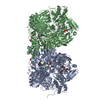
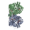
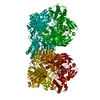
| Title | Xanthine Dehydrogenase from Bovine Milk with Inhibitor TEI-6720 Bound | ||||||
 Components Components | Xanthine Dehydrogenase | ||||||
 Keywords Keywords | OXIDOREDUCTASE / Molybdopterin / TEI-6720 / Flavoprotein / FAD / Iron-Sulfur Center / Fe2/S2 Center | ||||||
| Function / homology |  Function and homology informationxanthine dehydrogenase complex / xanthine dehydrogenase / xanthine oxidase / xanthine oxidase activity / xanthine catabolic process / xanthine dehydrogenase activity / molybdenum ion binding / molybdopterin cofactor binding / FAD binding / 2 iron, 2 sulfur cluster binding ...xanthine dehydrogenase complex / xanthine dehydrogenase / xanthine oxidase / xanthine oxidase activity / xanthine catabolic process / xanthine dehydrogenase activity / molybdenum ion binding / molybdopterin cofactor binding / FAD binding / 2 iron, 2 sulfur cluster binding / peroxisome / flavin adenine dinucleotide binding / iron ion binding / protein homodimerization activity / extracellular space Function and homology informationxanthine dehydrogenase complex / xanthine dehydrogenase / xanthine oxidase / xanthine oxidase activity / xanthine catabolic process / xanthine dehydrogenase activity / molybdenum ion binding / molybdopterin cofactor binding / FAD binding / 2 iron, 2 sulfur cluster binding ...xanthine dehydrogenase complex / xanthine dehydrogenase / xanthine oxidase / xanthine oxidase activity / xanthine catabolic process / xanthine dehydrogenase activity / molybdenum ion binding / molybdopterin cofactor binding / FAD binding / 2 iron, 2 sulfur cluster binding / peroxisome / flavin adenine dinucleotide binding / iron ion binding / protein homodimerization activity / extracellular spaceSimilarity search - Function | ||||||
| Biological species |  Bos taurus (cattle) Bos taurus (cattle) | ||||||
| Method | X-RAY DIFFRACTION / SYNCHROTRON / MOLECULAR REPLACEMENT / Resolution: 2.8 Å | ||||||
 Authors Authors | Okamoto, K. / Eger, B.T. / Nishino, T. / Kondo, S. / Pai, E.F. / Nishino, T. | ||||||
 Citation Citation | Journal: J.BIOL.CHEM. / Year: 2003 Title: An Extremely Potent Inhibitor of Xanthine Oxidoreductase: Crystal Structure of the Enzyme-Inhibitor Complex and Mechanism of Inhibition Authors: Okamoto, K. / Eger, B.T. / Nishino, T. / Kondo, S. / Pai, E.F. / Nishino, T. #1: Journal: EUR.J.PHARMACOL. / Year: 1993Title: Hypouricemic effect of the novel xanthine oxidase inhibitor, TEI-6720, in rodents Authors: Osada, Y. / Tsuchimoto, M. / Fukushima, H. / Takahashi, K. / Kondo, S. / Hasegawa, M. / Komoriya, K. | ||||||
| History |
|
- Structure visualization
| Structure viewer | Molecule: MolmilJmol/JSmol |
|---|
- Downloads & links
-Download
| PDBx/mmCIF format | 1n5x.cif.gz | 449.3 KB | Display | PDBx/mmCIF format |
|---|---|---|---|---|
| PDB format | pdb1n5x.ent.gz | 350.3 KB | Display | PDB format |
| PDBx/mmJSON format | 1n5x.json.gz | Tree view | PDBx/mmJSON format | |
| Others |  Other downloads Other downloads |
-Validation report
| Arichive directory | https://data.pdbj.org/pub/pdb/validation_reports/n5/1n5xftp://data.pdbj.org/pub/pdb/validation_reports/n5/1n5x | HTTPS FTP |
|---|
-Related structure data
| Related structure data | 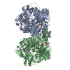 1fo4S S: Starting model for refinement |
|---|---|
| Similar structure data |


-Links
 PDBj
PDBj


- Assembly
| Deposited unit | 
| ||||||||||||
|---|---|---|---|---|---|---|---|---|---|---|---|---|---|
| 1 |
| ||||||||||||
| Unit cell |
| ||||||||||||
| Noncrystallographic symmetry (NCS) | NCS oper:
|
-Components
-Protein , 1 types, 2 molecules AB
| #1: Protein | Mass: 146861.953 Da / Num. of mol.: 2 / Source method: isolated from a natural source / Source: (natural) Bos taurus (cattle) / Secretion: milk / References: UniProt: P80457, 1.1.1.204 |
|---|
-Non-polymers , 5 types, 12 molecules 


| #2: Chemical | ChemComp-FES / Iron–sulfur cluster Mass: 175.820 Da / Num. of mol.: 4 / Source method: obtained synthetically / Formula: Fe2S2 Mass: 175.820 Da / Num. of mol.: 4 / Source method: obtained synthetically / Formula: Fe2S2#3: Chemical |  Mass: 395.352 Da / Num. of mol.: 2 / Source method: obtained synthetically / Formula: C10H14N5O6PS2 Mass: 395.352 Da / Num. of mol.: 2 / Source method: obtained synthetically / Formula: C10H14N5O6PS2#4: Chemical |  Mass: 161.012 Da / Num. of mol.: 2 / Source method: obtained synthetically / Formula: HMoO2S Mass: 161.012 Da / Num. of mol.: 2 / Source method: obtained synthetically / Formula: HMoO2S#5: Chemical | Flavin adenine dinucleotide Mass: 785.550 Da / Num. of mol.: 2 / Source method: obtained synthetically / Formula: C27H33N9O15P2 / Comment: FAD*YM Mass: 785.550 Da / Num. of mol.: 2 / Source method: obtained synthetically / Formula: C27H33N9O15P2 / Comment: FAD*YM#6: Chemical | Febuxostat Mass: 316.375 Da / Num. of mol.: 2 / Source method: obtained synthetically / Formula: C16H16N2O3S / Comment: medication*YM Mass: 316.375 Da / Num. of mol.: 2 / Source method: obtained synthetically / Formula: C16H16N2O3S / Comment: medication*YM |
|---|
-Experimental details
-Experiment
| Experiment | Method: X-RAY DIFFRACTION / Number of used crystals: 1 |
|---|
- Sample preparation
| Crystal | Density Matthews: 2.63 Å3/Da / Density % sol: 53.21 % | ||||||||||||||||||||||||||||||||||||||||||||||||||||||
|---|---|---|---|---|---|---|---|---|---|---|---|---|---|---|---|---|---|---|---|---|---|---|---|---|---|---|---|---|---|---|---|---|---|---|---|---|---|---|---|---|---|---|---|---|---|---|---|---|---|---|---|---|---|---|---|
| Crystal grow | Temperature: 293 K / Method: batch slide Details: PEG4000, DTT, Glycerol, Batch Slide, temperature 293K | ||||||||||||||||||||||||||||||||||||||||||||||||||||||
| Crystal grow | *PLUS Temperature: 20 ℃ / Method: unknown | ||||||||||||||||||||||||||||||||||||||||||||||||||||||
| Components of the solutions | *PLUS
|
-Data collection
| Diffraction | Mean temperature: 100 K |
|---|---|
| Diffraction source | Source: SYNCHROTRON / Site: SPring-8  / Beamline: BL40B2 / Wavelength: 1 Å / Beamline: BL40B2 / Wavelength: 1 Å |
| Detector | Type: ADSC QUANTUM 4r / Detector: CCD / Date: Mar 22, 2002 |
| Radiation | Protocol: SINGLE WAVELENGTH / Monochromatic (M) / Laue (L): M / Scattering type: x-ray |
| Radiation wavelength | Wavelength: 1 Å / Relative weight: 1 |
| Reflection | Resolution: 2.8→20 Å / Num. all: 78047 / Num. obs: 74031 / % possible obs: 99.1 % / Observed criterion σ(I): -3 / Redundancy: 3.24 % / Biso Wilson estimate: 21 Å2 / Rmerge(I) obs: 0.111 / Rsym value: 0.111 / Net I/σ(I): 9.59 |
| Reflection shell | Resolution: 2.8→2.85 Å / Redundancy: 2.84 % / Rmerge(I) obs: 0.442 / Mean I/σ(I) obs: 2.17 / Num. unique all: 3646 / Rsym value: 0.442 / % possible all: 98.2 |
| Reflection | *PLUS Lowest resolution: 20 Å / % possible obs: 99 % / Rmerge(I) obs: 0.116 |
| Reflection shell | *PLUS Highest resolution: 2.8 Å / % possible obs: 98.3 % / Rmerge(I) obs: 0.438 / Mean I/σ(I) obs: 2.2 |
- Processing
| Software |
| |||||||||||||||||||||||||
|---|---|---|---|---|---|---|---|---|---|---|---|---|---|---|---|---|---|---|---|---|---|---|---|---|---|---|
| Refinement | Method to determine structure: MOLECULAR REPLACEMENT Starting model: PDB 1FO4 Resolution: 2.8→20 Å / Isotropic thermal model: isotropic / Cross valid method: THROUGHOUT / σ(F): 0 / Stereochemistry target values: Engh & Huber / Details: STRICT NCS WAS USED THROUGHOUT
| |||||||||||||||||||||||||
| Displacement parameters | Biso mean: 21 Å2 | |||||||||||||||||||||||||
| Refine analyze |
| |||||||||||||||||||||||||
| Refinement step | Cycle: LAST / Resolution: 2.8→20 Å
| |||||||||||||||||||||||||
| Refine LS restraints |
| |||||||||||||||||||||||||
| Refine LS restraints NCS | NCS model details: STRICT NCS | |||||||||||||||||||||||||
| LS refinement shell | Resolution: 2.8→2.85 Å / Rfactor Rfree error: 0.038
| |||||||||||||||||||||||||
| Refinement | *PLUS Highest resolution: 2.8 Å / Lowest resolution: 20 Å / Rfactor obs: 0.244 | |||||||||||||||||||||||||
| Solvent computation | *PLUS | |||||||||||||||||||||||||
| Displacement parameters | *PLUS Biso mean: 21 Å2 | |||||||||||||||||||||||||
| Refine LS restraints | *PLUS
| |||||||||||||||||||||||||
| LS refinement shell | *PLUS Highest resolution: 2.8 Å |
