 Movie
Movie Controller


+ Open data

- Basic information
| Entry | Database: PDB / ID: 1n4l | ||||||
|---|---|---|---|---|---|---|---|
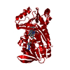
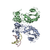
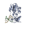
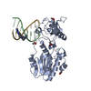
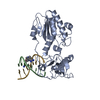
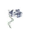
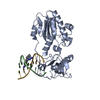
| Title | A DNA analogue of the polypurine tract of HIV-1 | ||||||
 Components Components |
| ||||||
 Keywords Keywords | TRANSFERASE/DNA / MMLV RT / Polypurine Tract /  HIV-1 / Asymmetric DNA / protein-DNA complex / TRANSFERASE-DNA COMPLEX HIV-1 / Asymmetric DNA / protein-DNA complex / TRANSFERASE-DNA COMPLEX | ||||||
| Function / homology |  Function and homology information Function and homology informationretroviral 3' processing activity / host cell late endosome membrane / DNA catabolic process / Hydrolases; Acting on peptide bonds (peptidases); Aspartic endopeptidases / ribonuclease H / virion assembly / protein-DNA complex / host multivesicular body / RNA-directed DNA polymerase / viral genome integration into host DNA ...retroviral 3' processing activity / host cell late endosome membrane / DNA catabolic process / Hydrolases; Acting on peptide bonds (peptidases); Aspartic endopeptidases / ribonuclease H / virion assembly / protein-DNA complex / host multivesicular body / RNA-directed DNA polymerase / viral genome integration into host DNA / establishment of integrated proviral latency / RNA-directed DNA polymerase activity / RNA-DNA hybrid ribonuclease activity / Transferases; Transferring phosphorus-containing groups; Nucleotidyltransferases / symbiont-mediated suppression of host gene expression / viral nucleocapsid / DNA recombination / structural constituent of virion / Hydrolases; Acting on ester bonds / DNA-directed DNA polymerase / aspartic-type endopeptidase activity / DNA-directed DNA polymerase activity / symbiont entry into host cell / host cell plasma membrane / proteolysis / DNA binding / RNA binding / zinc ion binding / membraneSimilarity search - Function | ||||||
| Biological species |  Moloney murine leukemia virus Moloney murine leukemia virus | ||||||
| Method | X-RAY DIFFRACTION / SYNCHROTRON / MOLECULAR REPLACEMENT / Resolution: 2 Å | ||||||
 Authors Authors | Cote, M.L. / Pflomm, M. / Georgiadis, M.M. | ||||||
 Citation Citation | Journal: J.Mol.Biol. / Year: 2003 Title: Staying Straight with A-tracts: A DNA Analog of the HIV-1 Polypurine Tract Authors: Cote, M.L. / Pflomm, M. / Georgiadis, M.M. #1: Journal: J.Mol.Biol. / Year: 2000Title: Crystal Structures of an N-terminal Fragment from Moloney Murine Leukemia Virus Reverse Transcriptase Complexed with Nucleic Acid: Functional Implications for Template-primer Binding to the Fingers Domain Authors: Najmudin, S. / Cote, M.L. / Sun, D. / Yohannan, S. / Montano, S.P. / Gu, J. / Georgiadis, M.M. #2: Journal: Acta Crystallogr.,Sect.D / Year: 2000Title: Use of an N-terminal fragment from Moloney murine leukemia virus reverse transcriptase to facilitate crystallization and analysis of a pseudo-16-mer DNA molecule containing G-A mispairs Authors: Cote, M.L. / Yohannan, S.J. / Georgiadis, M.M. #3: Journal: Acta Crystallogr.,Sect.D / Year: 2001Title: Structure of a pseudo-16-mer DNA with stacked guanines and two G-A mispairs coomplexed with the N-terminal fragment of Moloney murine leukemia virus reverse transcriptase Authors: Cote, M. / Georgaidis, M.M. | ||||||
| History |
|
- Structure visualization
| Structure viewer | Molecule: MolmilJmol/JSmol |
|---|
- Downloads & links
-Download
| PDBx/mmCIF format | 1n4l.cif.gz | 87.5 KB | Display | PDBx/mmCIF format |
|---|---|---|---|---|
| PDB format | pdb1n4l.ent.gz | 61.6 KB | Display | PDB format |
| PDBx/mmJSON format | 1n4l.json.gz | Tree view | PDBx/mmJSON format | |
| Others |  Other downloads Other downloads |
-Validation report
| Arichive directory | https://data.pdbj.org/pub/pdb/validation_reports/n4/1n4lftp://data.pdbj.org/pub/pdb/validation_reports/n4/1n4l | HTTPS FTP |
|---|
-Related structure data
| Related structure data | 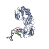 1d1uS  1n85 S: Starting model for refinement |
|---|---|
| Similar structure data |


-Links
 PDBj
PDBj


- Assembly
| Deposited unit | 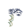
| ||||||||||
|---|---|---|---|---|---|---|---|---|---|---|---|
| 1 | 
| ||||||||||
| Unit cell |
|
-Components
| #1: DNA chain | Mass: 4929.254 Da / Num. of mol.: 1 / Source method: obtained synthetically |
|---|---|
| #2: DNA chain | Mass: 4862.188 Da / Num. of mol.: 1 / Source method: obtained synthetically |
| #3: Protein | / E.C.2.7.7.49 / MMLV RT Mass: 28934.287 Da / Num. of mol.: 1 Source method: isolated from a genetically manipulated source Details: part of Pol polyprotein / Source: (gene. exp.) Moloney murine leukemia virus / Genus: Gammaretrovirus / Species: Murine leukemia virus / Plasmid: pET15b / Species (production host): Escherichia coli / Production host:  Escherichia coli BL21 (bacteria) / Strain (production host): BL21 / References: UniProt: P03355, RNA-directed DNA polymerase Escherichia coli BL21 (bacteria) / Strain (production host): BL21 / References: UniProt: P03355, RNA-directed DNA polymerase |
| #4: Water | ChemComp-HOH / Water Mass: 18.015 Da / Num. of mol.: 145 / Source method: isolated from a natural source / Formula: H2O Mass: 18.015 Da / Num. of mol.: 145 / Source method: isolated from a natural source / Formula: H2O |
-Experimental details
-Experiment
| Experiment | Method: X-RAY DIFFRACTION / Number of used crystals: 2 |
|---|
- Sample preparation
| Crystal | Density Matthews: 2.35 Å3/Da / Density % sol: 47.1 % | ||||||||||||||||||||||||||||||||||||||||||||||||||||||||||||||||||
|---|---|---|---|---|---|---|---|---|---|---|---|---|---|---|---|---|---|---|---|---|---|---|---|---|---|---|---|---|---|---|---|---|---|---|---|---|---|---|---|---|---|---|---|---|---|---|---|---|---|---|---|---|---|---|---|---|---|---|---|---|---|---|---|---|---|---|---|
| Crystal grow | Temperature: 277 K / Method: vapor diffusion, hanging drop / pH: 6.5 Details: PEG 4000, NaCl, ADA, MgCl2, HEPES, pH 6.5, VAPOR DIFFUSION, HANGING DROP at 277K | ||||||||||||||||||||||||||||||||||||||||||||||||||||||||||||||||||
| Components of the solutions |
| ||||||||||||||||||||||||||||||||||||||||||||||||||||||||||||||||||
| Crystal grow | *PLUS Temperature: 4 ℃ / Method: vapor diffusion, hanging drop | ||||||||||||||||||||||||||||||||||||||||||||||||||||||||||||||||||
| Components of the solutions | *PLUS
|
-Data collection
| Diffraction |
| ||||||||||||||||||
|---|---|---|---|---|---|---|---|---|---|---|---|---|---|---|---|---|---|---|---|
| Diffraction source |
| ||||||||||||||||||
| Detector |
| ||||||||||||||||||
| Radiation |
| ||||||||||||||||||
| Radiation wavelength |
| ||||||||||||||||||
| Reflection | Resolution: 2→50 Å / Num. all: 24798 / Num. obs: 23122 / % possible obs: 93.2 % / Observed criterion σ(I): -3 | ||||||||||||||||||
| Reflection shell | Resolution: 2→2.1 Å / Redundancy: 3.1 % / Mean I/σ(I) obs: 5.8 / Num. unique all: 2691 / Rsym value: 0.15 / % possible all: 76.1 | ||||||||||||||||||
| Reflection | *PLUS Highest resolution: 2 Å / Lowest resolution: 20 Å / % possible obs: 93.9 % / Rmerge(I) obs: 0.118 | ||||||||||||||||||
| Reflection shell | *PLUS Highest resolution: 2 Å / Lowest resolution: 2.07 Å / % possible obs: 84.8 % / Mean I/σ(I) obs: 5.7 |

- Processing
| Software |
| |||||||||||||||||||||||||
|---|---|---|---|---|---|---|---|---|---|---|---|---|---|---|---|---|---|---|---|---|---|---|---|---|---|---|
| Refinement | Method to determine structure: MOLECULAR REPLACEMENT Starting model: PDB ENTRY 1D1U Resolution: 2→50 Å / Isotropic thermal model: Isotropic / Cross valid method: THROUGHOUT / σ(F): 0 / Stereochemistry target values: Engh & Huber
| |||||||||||||||||||||||||
| Displacement parameters | Biso mean: 42.8 Å2 | |||||||||||||||||||||||||
| Refine analyze | Luzzati coordinate error obs: 0.29 Å / Luzzati sigma a obs: 0.2 Å | |||||||||||||||||||||||||
| Refinement step | Cycle: LAST / Resolution: 2→50 Å
| |||||||||||||||||||||||||
| Refine LS restraints |
| |||||||||||||||||||||||||
| LS refinement shell | Resolution: 2→2.1 Å
| |||||||||||||||||||||||||
| Refinement | *PLUS Highest resolution: 2 Å / Lowest resolution: 50 Å / Rfactor Rwork: 0.24 | |||||||||||||||||||||||||
| Solvent computation | *PLUS | |||||||||||||||||||||||||
| Displacement parameters | *PLUS | |||||||||||||||||||||||||
| Refine LS restraints | *PLUS
|
