 Movie
Movie Controller


+ Open data

- Basic information
| Entry | Database: PDB / ID: 1n47 | |||||||||
|---|---|---|---|---|---|---|---|---|---|---|
| Title | Isolectin B4 from Vicia villosa in complex with the Tn antigen | |||||||||
 Components Components | Isolectin B4 | |||||||||
 Keywords Keywords | SUGAR BINDING PROTEIN / cancer antigen / Vicia villosa lectin / glycoprotein Tn-binding protein / carbohydrate recognition | |||||||||
| Function / homology |  Function and homology information Function and homology information | |||||||||
| Biological species |   Vicia villosa (hairy vetch) Vicia villosa (hairy vetch) | |||||||||
| Method | X-RAY DIFFRACTION / SYNCHROTRON / MOLECULAR REPLACEMENT / Resolution: 2.7 Å | |||||||||
 Authors Authors | Babino, A. / Tello, D. / Rojas, A. / Bay, S. / Osinaga, E. / Alzari, P.M. | |||||||||
 Citation Citation | Journal: FEBS Lett. / Year: 2003 Title: The crystal structure of a plant lectin in complex with the Tn antigen Authors: Babino, A. / Tello, D. / Rojas, A. / Bay, S. / Osinaga, E. / Alzari, P.M. #1: Journal: FEBS Lett. / Year: 1997Title: Amino acid sequence and three-dimensional structure of the Tn-specific isolectin B4 from Vicia villosa. Authors: Osinaga, E. / Tello, D. / Batthyany, C. / Bianchet, M. / Tavares, G. / Duran, R. / Cervenansky, C. / Camoin, L. / Roseto, A. / Alzari, P.M. | |||||||||
| History |
| |||||||||
| Remark 999 | sequence The author maintains that the sequence for the exact isoform crystallized here has not yet ...sequence The author maintains that the sequence for the exact isoform crystallized here has not yet been deposited in any sequence database. |
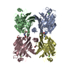
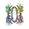
- Structure visualization
| Structure viewer | Molecule: MolmilJmol/JSmol |
|---|
- Downloads & links
-Download
| PDBx/mmCIF format | 1n47.cif.gz | 188.3 KB | Display | PDBx/mmCIF format |
|---|---|---|---|---|
| PDB format | pdb1n47.ent.gz | 151.8 KB | Display | PDB format |
| PDBx/mmJSON format | 1n47.json.gz | Tree view | PDBx/mmJSON format | |
| Others |  Other downloads Other downloads |
-Validation report
| Arichive directory | https://data.pdbj.org/pub/pdb/validation_reports/n4/1n47ftp://data.pdbj.org/pub/pdb/validation_reports/n4/1n47 | HTTPS FTP |
|---|
-Related structure data
| Similar structure data |
|---|


-Links
 PDBj
PDBj

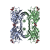
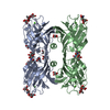
- Assembly
| Deposited unit | 
| ||||||||
|---|---|---|---|---|---|---|---|---|---|
| 1 |
| ||||||||
| Unit cell |
|
-Components
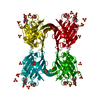
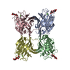
-Protein , 1 types, 4 molecules ABCD
| #1: Protein | Mass: 24710.346 Da / Num. of mol.: 4 / Source method: isolated from a natural source / Details: extracted from seeds / Source: (natural) Vicia villosa (hairy vetch) / Tissue: seed / References: UniProt: P56625 |
|---|
-Sugars , 2 types, 8 molecules 
| #2: Polysaccharide | alpha-L-fucopyranose-(1-3)-[2-acetamido-2-deoxy-beta-D-glucopyranose-(1-4)]2-acetamido-2-deoxy-beta- ...alpha-L-fucopyranose-(1-3)-[2-acetamido-2-deoxy-beta-D-glucopyranose-(1-4)]2-acetamido-2-deoxy-beta-D-glucopyranose / Mass: 570.542 Da / Num. of mol.: 4Source method: isolated from a genetically manipulated source #6: Sugar | ChemComp-A2G / N-Acetylgalactosamine Type: D-saccharide, alpha linking / Mass: 221.208 Da / Num. of mol.: 4 Type: D-saccharide, alpha linking / Mass: 221.208 Da / Num. of mol.: 4Source method: isolated from a genetically manipulated source Formula: C8H15NO6 |
|---|
-Non-polymers , 4 types, 69 molecules 


| #3: Chemical | ChemComp-CA /  Mass: 40.078 Da / Num. of mol.: 4 / Source method: obtained synthetically / Formula: Ca Mass: 40.078 Da / Num. of mol.: 4 / Source method: obtained synthetically / Formula: Ca#4: Chemical | ChemComp-MN /  Mass: 54.938 Da / Num. of mol.: 4 / Source method: obtained synthetically / Formula: Mn Mass: 54.938 Da / Num. of mol.: 4 / Source method: obtained synthetically / Formula: Mn#5: Chemical | ChemComp-SER / Serine Type: L-peptide linking / Mass: 105.093 Da / Num. of mol.: 4 / Source method: obtained synthetically / Formula: C3H7NO3 Type: L-peptide linking / Mass: 105.093 Da / Num. of mol.: 4 / Source method: obtained synthetically / Formula: C3H7NO3#7: Water | ChemComp-HOH / | WaterMass: 18.015 Da / Num. of mol.: 57 / Source method: isolated from a natural source / Formula: H2O |
|---|
-Experimental details
-Experiment
| Experiment | Method: X-RAY DIFFRACTION / Number of used crystals: 1 |
|---|
- Sample preparation
| Crystal | Density Matthews: 2.84 Å3/Da / Density % sol: 56.69 % | ||||||||||||||||||
|---|---|---|---|---|---|---|---|---|---|---|---|---|---|---|---|---|---|---|---|
| Crystal grow | Temperature: 291 K / Method: vapor diffusion, hanging drop / pH: 4.7 Details: acetate, MPD , pH 4.7, VAPOR DIFFUSION, HANGING DROP, temperature 291K | ||||||||||||||||||
| Crystal grow | *PLUS | ||||||||||||||||||
| Components of the solutions | *PLUS
|
-Data collection
| Diffraction | Mean temperature: 277 K |
|---|---|
| Diffraction source | Source: SYNCHROTRON / Site: LURE  / Beamline: DW32 / Wavelength: 0.93 Å / Beamline: DW32 / Wavelength: 0.93 Å |
| Detector | Type: MARRESEARCH / Detector: AREA DETECTOR |
| Radiation | Protocol: SINGLE WAVELENGTH / Monochromatic (M) / Laue (L): M / Scattering type: x-ray |
| Radiation wavelength | Wavelength: 0.93 Å / Relative weight: 1 |
| Reflection | Resolution: 2.7→20 Å / Num. obs: 30037 / % possible obs: 99.8 % / Observed criterion σ(F): 0 / Observed criterion σ(I): 0 / Redundancy: 4.7 % / Rsym value: 0.107 |
| Reflection shell | Resolution: 2.7→2.8 Å / Rsym value: 0.46 / % possible all: 99.8 |
| Reflection | *PLUS Num. measured all: 144637 / Rmerge(I) obs: 0.107 |
| Reflection shell | *PLUS Lowest resolution: 2.78 Å / % possible obs: 99.8 % / Rmerge(I) obs: 0.467 |
- Processing
| Software |
| |||||||||||||||||||||||||||||||||||||||||||||||||||||||||||||||||||||||||||
|---|---|---|---|---|---|---|---|---|---|---|---|---|---|---|---|---|---|---|---|---|---|---|---|---|---|---|---|---|---|---|---|---|---|---|---|---|---|---|---|---|---|---|---|---|---|---|---|---|---|---|---|---|---|---|---|---|---|---|---|---|---|---|---|---|---|---|---|---|---|---|---|---|---|---|---|---|
| Refinement | Method to determine structure: MOLECULAR REPLACEMENT Starting model: structure of unliganded lectin (from reference 1) Resolution: 2.7→20 Å / Cor.coef. Fo:Fc: 0.94 / Cor.coef. Fo:Fc free: 0.91 / SU B: 12.227 / SU ML: 0.251 / Cross valid method: THROUGHOUT / σ(F): 0 / ESU R Free: 0.329
| |||||||||||||||||||||||||||||||||||||||||||||||||||||||||||||||||||||||||||
| Solvent computation | Ion probe radii: 0.8 Å / Shrinkage radii: 0.8 Å / VDW probe radii: 1.4 Å / Solvent model: BABINET MODEL WITH MASK | |||||||||||||||||||||||||||||||||||||||||||||||||||||||||||||||||||||||||||
| Displacement parameters | Biso mean: 33.566 Å2
| |||||||||||||||||||||||||||||||||||||||||||||||||||||||||||||||||||||||||||
| Refinement step | Cycle: LAST / Resolution: 2.7→20 Å
| |||||||||||||||||||||||||||||||||||||||||||||||||||||||||||||||||||||||||||
| Refine LS restraints |
| |||||||||||||||||||||||||||||||||||||||||||||||||||||||||||||||||||||||||||
| LS refinement shell | Resolution: 2.7→2.769 Å / Total num. of bins used: 20 /
| |||||||||||||||||||||||||||||||||||||||||||||||||||||||||||||||||||||||||||
| Refinement | *PLUS Highest resolution: 2.7 Å / Lowest resolution: 20 Å / % reflection Rfree: 5 % / Rfactor Rfree: 0.238 / Rfactor Rwork: 0.188 | |||||||||||||||||||||||||||||||||||||||||||||||||||||||||||||||||||||||||||
| Solvent computation | *PLUS | |||||||||||||||||||||||||||||||||||||||||||||||||||||||||||||||||||||||||||
| Displacement parameters | *PLUS | |||||||||||||||||||||||||||||||||||||||||||||||||||||||||||||||||||||||||||
| Refine LS restraints | *PLUS
|
