 Movie
Movie Controller

[English] 日本語
 Yorodumi
- PDB-1n3z: Crystal structure of the [S-carboxyamidomethyl-Cys31, S-carboxyam... -
+ Open data

- Basic information
| Entry | Database: PDB / ID: 1n3z | ||||||
|---|---|---|---|---|---|---|---|
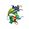
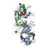
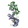
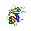
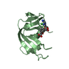
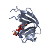
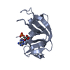
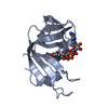
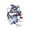
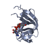
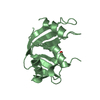
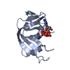
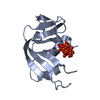
| Title | Crystal structure of the [S-carboxyamidomethyl-Cys31, S-carboxyamidomethyl-Cys32] monomeric derivative of the bovine seminal ribonuclease in the liganded state | ||||||
 Components Components | Ribonuclease, seminal | ||||||
 Keywords Keywords | HYDROLASE / Protein-nucleotide complex | ||||||
| Function / homology |  Function and homology informationpancreatic ribonuclease / ribonuclease A activity / RNA nuclease activity / metabolic process / nucleic acid binding / lyase activity / defense response to Gram-positive bacterium / extracellular region / identical protein binding Function and homology informationpancreatic ribonuclease / ribonuclease A activity / RNA nuclease activity / metabolic process / nucleic acid binding / lyase activity / defense response to Gram-positive bacterium / extracellular region / identical protein bindingSimilarity search - Function | ||||||
| Biological species |  Bos taurus (cattle) Bos taurus (cattle) | ||||||
| Method | X-RAY DIFFRACTION / SYNCHROTRON / MOLECULAR REPLACEMENT / Resolution: 1.65 Å | ||||||
 Authors Authors | Sica, F. / Di Fiore, A. / Zagari, A. / Mazzarella, L. | ||||||
 Citation Citation | Journal: Proteins / Year: 2003 Title: The unswapped chain of bovine seminal ribonuclease: Crystal structure of the free and liganded monomeric derivative Authors: Sica, F. / Di Fiore, A. / Zagari, A. / Mazzarella, L. #1: Journal: Acta Crystallogr.,Sect.D / Year: 1993Title: Bovine seminal ribonuclease: structure at 1.9 resolution Authors: Mazzarella, L. / Capasso, S. / Demasi, D. / Di Lorenzo, G. / Mattia, C.A. / Zagari, A. #2: Journal: Proc.Natl.Acad.Sci.USA / Year: 1995Title: Swapping structural determinants of ribonucleases: an energetic analysis of the hinge peptide 16-22 Authors: Mazzarella, L. / Vitagliano, L. / Zagari, A. #3: Journal: J.CRYST.GROWTH / Year: 1999Title: Crystallization of multiple forms of bovine seminal ribonuclease in the liganded and unliganded state Authors: Sica, F. / Adinolfi, S. / Berisio, R. / De Lorenzo, C. / Mazzarella, L. / Piccoli, R. / Vitagliano, L. / Zagari, A. #4: Journal: Protein Sci. / Year: 1998Title: Binding of a substrate analog to a domain swapping protein: X-ray structure of the complex of bovine seminal ribonuclease with uridylyl(2',5')adenosine Authors: Vitagliano, L. / Adinolfi, S. / Riccio, A. / Sica, F. / Zagari, A. / Mazzarella, L. #5: Journal: Biochemistry / Year: 1998Title: Coulombic effects of remote subsites on the active site of ribonuclease A Authors: Fisher, B.M. / Schultz, L.W. / Raines, R.T. | ||||||
| History |
|
- Structure visualization
| Structure viewer | Molecule: MolmilJmol/JSmol |
|---|
- Downloads & links
-Download
| PDBx/mmCIF format | 1n3z.cif.gz | 37.1 KB | Display | PDBx/mmCIF format |
|---|---|---|---|---|
| PDB format | pdb1n3z.ent.gz | 28.6 KB | Display | PDB format |
| PDBx/mmJSON format | 1n3z.json.gz | Tree view | PDBx/mmJSON format | |
| Others |  Other downloads Other downloads |
-Validation report
| Arichive directory | https://data.pdbj.org/pub/pdb/validation_reports/n3/1n3zftp://data.pdbj.org/pub/pdb/validation_reports/n3/1n3z | HTTPS FTP |
|---|
-Related structure data

-Links
 PDBj
PDBj


- Assembly
| Deposited unit | 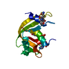
| ||||||||
|---|---|---|---|---|---|---|---|---|---|
| 1 |
| ||||||||
| Unit cell |
|
-Components
| #1: Protein | / Seminal RNase / Ribonuclease BS-1 Mass: 13746.742 Da / Num. of mol.: 1 / Source method: isolated from a natural source / Source: (natural) Bos taurus (cattle) / References: UniProt: P00669, EC: 3.1.27.5 |
|---|---|
| #2: Chemical | ChemComp-U3P /   Mass: 324.181 Da / Num. of mol.: 1 / Source method: obtained synthetically / Formula: C9H13N2O9P Mass: 324.181 Da / Num. of mol.: 1 / Source method: obtained synthetically / Formula: C9H13N2O9P |
| #3: Chemical | ChemComp-ADN / Adenosine  Mass: 267.241 Da / Num. of mol.: 1 / Source method: obtained synthetically / Formula: C10H13N5O4 Mass: 267.241 Da / Num. of mol.: 1 / Source method: obtained synthetically / Formula: C10H13N5O4 |
| #4: Water | ChemComp-HOH / Water Mass: 18.015 Da / Num. of mol.: 149 / Source method: isolated from a natural source / Formula: H2O Mass: 18.015 Da / Num. of mol.: 149 / Source method: isolated from a natural source / Formula: H2O |
-Experimental details
-Experiment
| Experiment | Method: X-RAY DIFFRACTION / Number of used crystals: 1 |
|---|
- Sample preparation
| Crystal | Density Matthews: 2.16 Å3/Da / Density % sol: 43.97 % | ||||||||||||||||||||||||||||||||||||
|---|---|---|---|---|---|---|---|---|---|---|---|---|---|---|---|---|---|---|---|---|---|---|---|---|---|---|---|---|---|---|---|---|---|---|---|---|---|
| Crystal grow | Temperature: 293 K / Method: vapor diffusion, sitting drop / pH: 8.7 Details: PEG 4000, sodium acetate, acetonitrile, glycerol (cryoprotectant), TRIS-HCl, pH 8.7, VAPOR DIFFUSION, SITTING DROP, temperature 293K | ||||||||||||||||||||||||||||||||||||
| Crystal grow | *PLUS Method: vapor diffusion, sitting drop | ||||||||||||||||||||||||||||||||||||
| Components of the solutions | *PLUS
|
-Data collection
| Diffraction | Mean temperature: 100 K |
|---|---|
| Diffraction source | Source: SYNCHROTRON / Site: ELETTRA  / Beamline: 5.2R / Wavelength: 1 Å / Beamline: 5.2R / Wavelength: 1 Å |
| Detector | Type: MARRESEARCH / Detector: IMAGE PLATE |
| Radiation | Protocol: SINGLE WAVELENGTH / Monochromatic (M) / Laue (L): M / Scattering type: x-ray |
| Radiation wavelength | Wavelength: 1 Å / Relative weight: 1 |
| Reflection | Resolution: 1.65→15 Å / Num. all: 14570 / Num. obs: 14570 / % possible obs: 99.9 % / Observed criterion σ(F): 0 / Observed criterion σ(I): 0 / Redundancy: 6 % / Biso Wilson estimate: 14.82 Å2 / Rmerge(I) obs: 0.037 / Net I/σ(I): 42 |
| Reflection shell | Resolution: 1.65→1.68 Å / Rmerge(I) obs: 0.098 / Mean I/σ(I) obs: 14 / Num. unique all: 723 / % possible all: 99.9 |
| Reflection | *PLUS Num. measured all: 99192 |
| Reflection shell | *PLUS % possible obs: 99.9 % |
- Processing
| Software |
| |||||||||||||||||||||||||
|---|---|---|---|---|---|---|---|---|---|---|---|---|---|---|---|---|---|---|---|---|---|---|---|---|---|---|
| Refinement | Method to determine structure: MOLECULAR REPLACEMENT / Resolution: 1.65→15 Å / Cross valid method: THROUGHOUT / σ(F): 2 / Stereochemistry target values: Engh & Huber
| |||||||||||||||||||||||||
| Displacement parameters | Biso mean: 16 Å2 | |||||||||||||||||||||||||
| Refinement step | Cycle: LAST / Resolution: 1.65→15 Å
| |||||||||||||||||||||||||
| Refine LS restraints |
| |||||||||||||||||||||||||
| LS refinement shell | Resolution: 1.65→1.68 Å
| |||||||||||||||||||||||||
| Refinement | *PLUS Rfactor Rfree: 0.23 | |||||||||||||||||||||||||
| Solvent computation | *PLUS | |||||||||||||||||||||||||
| Displacement parameters | *PLUS | |||||||||||||||||||||||||
| Refine LS restraints | *PLUS
|
