 Movie
Movie Controller

[English] 日本語
 Yorodumi
- PDB-1n3a: Structural and biochemical exploration of a critical amino acid i... -
+ Open data

- Basic information
| Entry | Database: PDB / ID: 1n3a | ||||||
|---|---|---|---|---|---|---|---|
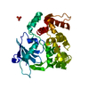
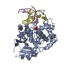
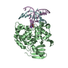
| Title | Structural and biochemical exploration of a critical amino acid in human 8-oxoguanine glycosylase | ||||||
 Components Components |
| ||||||
 Keywords Keywords |  hydrolase / lyase/DNA / HhH-GPD / glycosylase / DNA-repair / oxoguanine / lyase-DNA COMPLEX hydrolase / lyase/DNA / HhH-GPD / glycosylase / DNA-repair / oxoguanine / lyase-DNA COMPLEX | ||||||
| Function / homology |  Function and homology information Function and homology informationDefective OGG1 Substrate Binding / Defective OGG1 Substrate Processing / Defective OGG1 Localization / depurination / negative regulation of double-strand break repair via single-strand annealing / oxidized purine nucleobase lesion DNA N-glycosylase activity / base-excision repair, AP site formation / depyrimidination / Displacement of DNA glycosylase by APEX1 / 8-oxo-7,8-dihydroguanine DNA N-glycosylase activity ...Defective OGG1 Substrate Binding / Defective OGG1 Substrate Processing / Defective OGG1 Localization / depurination / negative regulation of double-strand break repair via single-strand annealing / oxidized purine nucleobase lesion DNA N-glycosylase activity / base-excision repair, AP site formation / depyrimidination / Displacement of DNA glycosylase by APEX1 / 8-oxo-7,8-dihydroguanine DNA N-glycosylase activity / response to folic acid / oxidized purine DNA binding / Hydrolases; Glycosylases; Hydrolysing N-glycosyl compounds / APEX1-Independent Resolution of AP Sites via the Single Nucleotide Replacement Pathway / response to light stimulus / Recognition and association of DNA glycosylase with site containing an affected purine / Cleavage of the damaged purine / Recognition and association of DNA glycosylase with site containing an affected pyrimidine / Cleavage of the damaged pyrimidine / cellular response to cadmium ion / class I DNA-(apurinic or apyrimidinic site) endonuclease activity / DNA-(apurinic or apyrimidinic site) lyase / nucleotide-excision repair / response to radiation / base-excision repair / nuclear matrix / response to estradiol / microtubule binding / endonuclease activity / response to ethanol / response to oxidative stress / damaged DNA binding / mitochondrial matrix / nuclear speck / response to xenobiotic stimulus / DNA damage response / regulation of DNA-templated transcription / negative regulation of apoptotic process / protein-containing complex / nucleoplasm / nucleus / cytosolSimilarity search - Function | ||||||
| Biological species |  Homo sapiens (human) Homo sapiens (human) | ||||||
| Method | X-RAY DIFFRACTION / SYNCHROTRON / MOLECULAR REPLACEMENT / Resolution: 2.2 Å | ||||||
 Authors Authors | Norman, D.P. / Chung, S.J. / Verdine, G.L. | ||||||
 Citation Citation | Journal: Biochemistry / Year: 2003 Title: Structural and biochemical exploration of a critical amino acid in human 8-oxoguanine glycosylase Authors: Norman, D.P. / Chung, S.J. / Verdine, G.L. | ||||||
| History |
|
- Structure visualization
| Structure viewer | Molecule: MolmilJmol/JSmol |
|---|
- Downloads & links
-Download
| PDBx/mmCIF format | 1n3a.cif.gz | 95.8 KB | Display | PDBx/mmCIF format |
|---|---|---|---|---|
| PDB format | pdb1n3a.ent.gz | 68.9 KB | Display | PDB format |
| PDBx/mmJSON format | 1n3a.json.gz | Tree view | PDBx/mmJSON format | |
| Others |  Other downloads Other downloads |
-Validation report
| Arichive directory | https://data.pdbj.org/pub/pdb/validation_reports/n3/1n3aftp://data.pdbj.org/pub/pdb/validation_reports/n3/1n3a | HTTPS FTP |
|---|
-Related structure data
| Related structure data |  1n39C 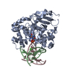 1n3cC  1fn7S S: Starting model for refinement C: citing same article ( |
|---|---|
| Similar structure data |


-Links
 PDBj
PDBj


- Assembly
| Deposited unit | 
| ||||||||
|---|---|---|---|---|---|---|---|---|---|
| 1 |
| ||||||||
| Unit cell |
|
-Components
| #1: DNA chain | Mass: 4635.010 Da / Num. of mol.: 1 / Source method: obtained synthetically |
|---|---|
| #2: DNA chain | Mass: 4396.838 Da / Num. of mol.: 1 / Source method: obtained synthetically |
| #3: Protein | Mass: 35661.348 Da / Num. of mol.: 1 / Mutation: D268Q Source method: isolated from a genetically manipulated source Source: (gene. exp.) Homo sapiens (human) / Gene: OGG1 / Plasmid: pet30a / Species (production host): Escherichia coli / Production host:  Escherichia coli BL21(DE3) (bacteria) / Strain (production host): BL21 DE3 Escherichia coli BL21(DE3) (bacteria) / Strain (production host): BL21 DE3References: UniProt: O15527, Hydrolases; Glycosylases; Hydrolysing N-glycosyl compounds, DNA-(apurinic or apyrimidinic site) lyase |
| #4: Chemical | ChemComp-CA /   Mass: 40.078 Da / Num. of mol.: 1 / Source method: obtained synthetically / Formula: Ca Mass: 40.078 Da / Num. of mol.: 1 / Source method: obtained synthetically / Formula: Ca |
| #5: Water | ChemComp-HOH / Water Mass: 18.015 Da / Num. of mol.: 148 / Source method: isolated from a natural source / Formula: H2O Mass: 18.015 Da / Num. of mol.: 148 / Source method: isolated from a natural source / Formula: H2O |
-Experimental details
-Experiment
| Experiment | Method: X-RAY DIFFRACTION / Number of used crystals: 1 |
|---|
- Sample preparation
| Crystal | Density Matthews: 2.88 Å3/Da / Density % sol: 57.36 % | ||||||||||||||||||||||||||||||||||||||||||||||||
|---|---|---|---|---|---|---|---|---|---|---|---|---|---|---|---|---|---|---|---|---|---|---|---|---|---|---|---|---|---|---|---|---|---|---|---|---|---|---|---|---|---|---|---|---|---|---|---|---|---|
| Crystal grow | Temperature: 277 K / Method: vapor diffusion, hanging drop / pH: 6.5 Details: cacodylate, calcium acetate, 16-18% PEG 8000, pH 6.5, VAPOR DIFFUSION, HANGING DROP, temperature 277K | ||||||||||||||||||||||||||||||||||||||||||||||||
| Components of the solutions |
| ||||||||||||||||||||||||||||||||||||||||||||||||
| Crystal grow | *PLUS Temperature: 4 ℃ / pH: 7.4 / Details: Bruner, S.D., (2000) Nature, 403, 859. | ||||||||||||||||||||||||||||||||||||||||||||||||
| Components of the solutions | *PLUS
|
-Data collection
| Diffraction | Mean temperature: 100 K |
|---|---|
| Diffraction source | Source: SYNCHROTRON / Site: CHESS  / Beamline: A1 / Wavelength: 0.93 Å / Beamline: A1 / Wavelength: 0.93 Å |
| Detector | Type: ADSC QUANTUM 4 / Detector: CCD / Date: Apr 9, 2001 |
| Radiation | Protocol: SINGLE WAVELENGTH / Monochromatic (M) / Laue (L): M / Scattering type: x-ray |
| Radiation wavelength | Wavelength: 0.93 Å / Relative weight: 1 |
| Reflection | Resolution: 2.2→30 Å / Num. all: 30819 / Num. obs: 29555 / % possible obs: 95.9 % / Observed criterion σ(F): 1 / Observed criterion σ(I): 1 / Redundancy: 5.1 % / Biso Wilson estimate: 28.3 Å2 / Rmerge(I) obs: 0.064 / Net I/σ(I): 23 |
| Reflection shell | Resolution: 2.2→2.34 Å / Rmerge(I) obs: 0.403 / Mean I/σ(I) obs: 4.7 / % possible all: 92.4 |
| Reflection | *PLUS Highest resolution: 2.2 Å / Lowest resolution: 30 Å / Num. obs: 29555 / % possible obs: 95.9 % / Num. measured all: 149944 / Rmerge(I) obs: 0.064 |
| Reflection shell | *PLUS Highest resolution: 2.2 Å / % possible obs: 92.4 % / Rmerge(I) obs: 0.403 / Mean I/σ(I) obs: 4.7 |
- Processing
| Software |
| ||||||||||||||||||||||||||||||||||||
|---|---|---|---|---|---|---|---|---|---|---|---|---|---|---|---|---|---|---|---|---|---|---|---|---|---|---|---|---|---|---|---|---|---|---|---|---|---|
| Refinement | Method to determine structure: MOLECULAR REPLACEMENT Starting model: 1FN7 Resolution: 2.2→29.93 Å / Rfactor Rfree error: 0.007 / Isotropic thermal model: RESTRAINED / Cross valid method: THROUGHOUT / σ(F): 0 / σ(I): 0 / Stereochemistry target values: Engh & Huber
| ||||||||||||||||||||||||||||||||||||
| Solvent computation | Solvent model: FLAT MODEL / Bsol: 42.7526 Å2 / ksol: 0.349783 e/Å3 | ||||||||||||||||||||||||||||||||||||
| Displacement parameters | Biso mean: 48.5 Å2
| ||||||||||||||||||||||||||||||||||||
| Refine analyze | Luzzati coordinate error free: 0.35 Å / Luzzati sigma a free: 0.33 Å | ||||||||||||||||||||||||||||||||||||
| Refinement step | Cycle: LAST / Resolution: 2.2→29.93 Å
| ||||||||||||||||||||||||||||||||||||
| Refine LS restraints |
| ||||||||||||||||||||||||||||||||||||
| LS refinement shell | Resolution: 2.2→2.34 Å / Rfactor Rfree error: 0.022 / Total num. of bins used: 6
| ||||||||||||||||||||||||||||||||||||
| Xplor file |
| ||||||||||||||||||||||||||||||||||||
| Refinement | *PLUS Lowest resolution: 30 Å / Rfactor Rfree: 0.27 | ||||||||||||||||||||||||||||||||||||
| Solvent computation | *PLUS | ||||||||||||||||||||||||||||||||||||
| Displacement parameters | *PLUS | ||||||||||||||||||||||||||||||||||||
| Refine LS restraints | *PLUS
| ||||||||||||||||||||||||||||||||||||
| LS refinement shell | *PLUS Rfactor Rfree: 0.34 / Rfactor Rwork: 0.33 |
