 Movie
Movie Controller

[English] 日本語
 Yorodumi
- PDB-1my6: The 1.6 A Structure of Fe-Superoxide Dismutase from the thermophi... -
+ Open data

- Basic information
| Entry | Database: PDB / ID: 1my6 | ||||||
|---|---|---|---|---|---|---|---|
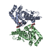
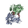
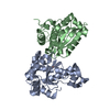
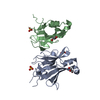
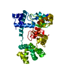
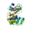
| Title | The 1.6 A Structure of Fe-Superoxide Dismutase from the thermophilic cyanobacterium Thermosynechococcus elongatus : Correlation of EPR and Structural Characteristics | ||||||
 Components Components | Iron (III) Superoxide Dismutase | ||||||
 Keywords Keywords |  OXIDOREDUCTASE / Iron Speroxide Dismutase / thermophile / reactive oxygen species / cyanobacteria / thermosynechococcus / Thermosynechococcus elongatus / Superoxide Dismutase / SOD / FeSOD / Iron(III) Superoxide Dismutase OXIDOREDUCTASE / Iron Speroxide Dismutase / thermophile / reactive oxygen species / cyanobacteria / thermosynechococcus / Thermosynechococcus elongatus / Superoxide Dismutase / SOD / FeSOD / Iron(III) Superoxide Dismutase | ||||||
| Function / homology |  Function and homology informationsuperoxide dismutase / superoxide dismutase activity / metal ion binding Function and homology informationsuperoxide dismutase / superoxide dismutase activity / metal ion bindingSimilarity search - Function | ||||||
| Biological species |   Thermosynechococcus elongatus (bacteria) Thermosynechococcus elongatus (bacteria) | ||||||
| Method | X-RAY DIFFRACTION / SYNCHROTRON / MOLECULAR REPLACEMENT / Resolution: 1.6 Å | ||||||
 Authors Authors | Yoshida, S.M. / Cascio, D. / Sawaya, M.R. / Yeates, T.O. / Kerfeld, C.A. | ||||||
 Citation Citation | Journal: J.BIOL.INORG.CHEM. / Year: 2003 Title: The 1.6 A resolution structure of Fe-superoxide dismutase from the thermophilic cyanobacterium Thermosynechococcus elongatus. Authors: Kerfeld, C.A. / Yoshida, S. / Tran, K.T. / Yeates, T.O. / Cascio, D. / Bottin, H. / Berthomieu, C. / Sugiura, M. / Boussac, A. | ||||||
| History |
|
- Structure visualization
| Structure viewer | Molecule: MolmilJmol/JSmol |
|---|
- Downloads & links
-Download
| PDBx/mmCIF format | 1my6.cif.gz | 184.4 KB | Display | PDBx/mmCIF format |
|---|---|---|---|---|
| PDB format | pdb1my6.ent.gz | 147.5 KB | Display | PDB format |
| PDBx/mmJSON format | 1my6.json.gz | Tree view | PDBx/mmJSON format | |
| Others |  Other downloads Other downloads |
-Validation report
| Arichive directory | https://data.pdbj.org/pub/pdb/validation_reports/my/1my6ftp://data.pdbj.org/pub/pdb/validation_reports/my/1my6 | HTTPS FTP |
|---|
-Related structure data
| Related structure data |  1iscS S: Starting model for refinement |
|---|---|
| Similar structure data |


-Links
 PDBj
PDBj
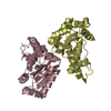
- Assembly
| Deposited unit | 
| ||||||||
|---|---|---|---|---|---|---|---|---|---|
| 1 |
| ||||||||
| Unit cell |
|
-Components
| #1: Protein | Mass: 22106.598 Da / Num. of mol.: 2 Source method: isolated from a genetically manipulated source Source: (gene. exp.) Thermosynechococcus elongatus (bacteria)Production host: Escherichia coli (E. coli) / References: UniProt: Q8DIR2, superoxide dismutase#2: Chemical | Iron  Mass: 55.845 Da / Num. of mol.: 2 / Source method: obtained synthetically / Formula: Fe Mass: 55.845 Da / Num. of mol.: 2 / Source method: obtained synthetically / Formula: Fe#3: Water | ChemComp-HOH / | Water Mass: 18.015 Da / Num. of mol.: 565 / Source method: isolated from a natural source / Formula: H2O Mass: 18.015 Da / Num. of mol.: 565 / Source method: isolated from a natural source / Formula: H2O |
|---|
-Experimental details
-Experiment
| Experiment | Method: X-RAY DIFFRACTION / Number of used crystals: 1 |
|---|
- Sample preparation
| Crystal | Density Matthews: 2.61 Å3/Da / Density % sol: 52.8 % | ||||||||||||||||||||||||||||||||||||
|---|---|---|---|---|---|---|---|---|---|---|---|---|---|---|---|---|---|---|---|---|---|---|---|---|---|---|---|---|---|---|---|---|---|---|---|---|---|
| Crystal grow | Temperature: 293.15 K / Method: vapor diffusion, hanging drop / pH: 6.7 Details: VAPOR DIFFUSION, HANGING DROP, 293.15K, 0.1M Sodium Cacodylate pH 6.7, 0.2M Magnesium Acetate Tetrahydrate, 20% PEG 6K | ||||||||||||||||||||||||||||||||||||
| Crystal grow | *PLUS pH: 7.5 / Method: vapor diffusion, hanging drop | ||||||||||||||||||||||||||||||||||||
| Components of the solutions | *PLUS
|
-Data collection
| Diffraction | Mean temperature: 100 K |
|---|---|
| Diffraction source | Source: SYNCHROTRON / Site: NSLS  / Beamline: X8C / Wavelength: 1.1 Å / Beamline: X8C / Wavelength: 1.1 Å |
| Detector | Type: ADSC QUANTUM 4 / Detector: CCD / Date: Jun 2, 2002 |
| Radiation | Protocol: SINGLE WAVELENGTH / Monochromatic (M) / Laue (L): M / Scattering type: x-ray |
| Radiation wavelength | Wavelength: 1.1 Å / Relative weight: 1 |
| Reflection | Resolution: 1.6→70 Å / Num. obs: 53851 / % possible obs: 94.5 % / Observed criterion σ(F): 0 / Observed criterion σ(I): -3 / Redundancy: 16.18 % / Biso Wilson estimate: 15.3 Å2 / Rsym value: 0.088 / Net I/σ(I): 6.35 |
| Reflection shell | Resolution: 1.6→1.642 Å / Redundancy: 14.28 % / Rmerge(I) obs: 0.32 / Mean I/σ(I) obs: 6.35 / Num. unique all: 5643 / Rsym value: 0.32 / % possible all: 93.4 |
| Reflection | *PLUS % possible obs: 99.57 % / Num. measured all: 274614 / Rmerge(I) obs: 0.088 |
| Reflection shell | *PLUS Lowest resolution: 1.66 Å / % possible obs: 94.5 % / Rmerge(I) obs: 0.322 |
- Processing
| Software |
| ||||||||||||||||||||||||||||||||||||||||||||||||||||||||||||||||||||||||||||||||||||||||||||||||||||||||||||||||||||||||||||||||||
|---|---|---|---|---|---|---|---|---|---|---|---|---|---|---|---|---|---|---|---|---|---|---|---|---|---|---|---|---|---|---|---|---|---|---|---|---|---|---|---|---|---|---|---|---|---|---|---|---|---|---|---|---|---|---|---|---|---|---|---|---|---|---|---|---|---|---|---|---|---|---|---|---|---|---|---|---|---|---|---|---|---|---|---|---|---|---|---|---|---|---|---|---|---|---|---|---|---|---|---|---|---|---|---|---|---|---|---|---|---|---|---|---|---|---|---|---|---|---|---|---|---|---|---|---|---|---|---|---|---|---|---|
| Refinement | Method to determine structure: MOLECULAR REPLACEMENT Starting model: FeSOD from E.coli, PDB entry 1isc Resolution: 1.6→70 Å / Cor.coef. Fo:Fc: 0.941 / Cor.coef. Fo:Fc free: 0.923 / SU B: 1.748 / SU ML: 0.065 / Cross valid method: THROUGHOUT / ESU R: 0.136 / ESU R Free: 0.101 / Stereochemistry target values: MAXIMUM LIKELIHOOD Details: HYDROGENS HAVE BEEN ADDED IN THE RIDING POSITIONS. Refined with anisotropic temperature factors. NCS restraints were not applied on the 2 molecules in the asymmetric unit.
| ||||||||||||||||||||||||||||||||||||||||||||||||||||||||||||||||||||||||||||||||||||||||||||||||||||||||||||||||||||||||||||||||||
| Solvent computation | Ion probe radii: 0.8 Å / Shrinkage radii: 0.8 Å / VDW probe radii: 1.4 Å / Solvent model: BABINET MODEL WITH MASK | ||||||||||||||||||||||||||||||||||||||||||||||||||||||||||||||||||||||||||||||||||||||||||||||||||||||||||||||||||||||||||||||||||
| Displacement parameters | Biso mean: 15.669 Å2
| ||||||||||||||||||||||||||||||||||||||||||||||||||||||||||||||||||||||||||||||||||||||||||||||||||||||||||||||||||||||||||||||||||
| Refinement step | Cycle: LAST / Resolution: 1.6→70 Å
| ||||||||||||||||||||||||||||||||||||||||||||||||||||||||||||||||||||||||||||||||||||||||||||||||||||||||||||||||||||||||||||||||||
| Refine LS restraints |
| ||||||||||||||||||||||||||||||||||||||||||||||||||||||||||||||||||||||||||||||||||||||||||||||||||||||||||||||||||||||||||||||||||
| LS refinement shell | Resolution: 1.6→1.642 Å / Total num. of bins used: 20 /
| ||||||||||||||||||||||||||||||||||||||||||||||||||||||||||||||||||||||||||||||||||||||||||||||||||||||||||||||||||||||||||||||||||
| Refinement | *PLUS % reflection Rfree: 5 % / Rfactor Rfree: 0.226 / Rfactor Rwork: 0.189 | ||||||||||||||||||||||||||||||||||||||||||||||||||||||||||||||||||||||||||||||||||||||||||||||||||||||||||||||||||||||||||||||||||
| Solvent computation | *PLUS | ||||||||||||||||||||||||||||||||||||||||||||||||||||||||||||||||||||||||||||||||||||||||||||||||||||||||||||||||||||||||||||||||||
| Displacement parameters | *PLUS | ||||||||||||||||||||||||||||||||||||||||||||||||||||||||||||||||||||||||||||||||||||||||||||||||||||||||||||||||||||||||||||||||||
| Refine LS restraints | *PLUS
|
