 Movie
Movie Controller

[English] 日本語
 Yorodumi
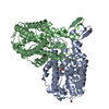
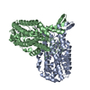
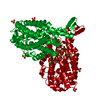
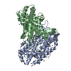
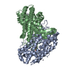
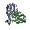
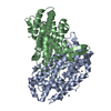
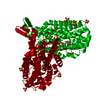
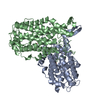
- PDB-1mxr: High resolution structure of Ribonucleotide reductase R2 from E. ... -
+ Open data

- Basic information
| Entry | Database: PDB / ID: 1mxr | ||||||
|---|---|---|---|---|---|---|---|
| Title | High resolution structure of Ribonucleotide reductase R2 from E. coli in its oxidised (Met) form | ||||||
 Components Components | Ribonucleotide reductase R2 | ||||||
 Keywords Keywords |  OXIDOREDUCTASE / Radical protein / di iron OXIDOREDUCTASE / Radical protein / di iron | ||||||
| Function / homology |  Function and homology information Function and homology informationribonucleoside diphosphate metabolic process / 2'-deoxyribonucleotide biosynthetic process / nucleobase-containing small molecule interconversion / ribonucleoside-diphosphate reductase complex / ribonucleoside-diphosphate reductase / ribonucleoside-diphosphate reductase activity, thioredoxin disulfide as acceptor / deoxyribonucleotide biosynthetic process / iron ion binding / identical protein binding / cytosol / cytoplasmSimilarity search - Function | ||||||
| Biological species |  Escherichia coli (E. coli) Escherichia coli (E. coli) | ||||||
| Method | X-RAY DIFFRACTION / SYNCHROTRON / MOLECULAR REPLACEMENT / Resolution: 1.42 Å | ||||||
 Authors Authors | Andersson, M.A. / Hogbom, M. / Nordlund, P. | ||||||
 Citation Citation | Journal: Proc.Natl.Acad.Sci.Usa / Year: 2003 Title: Displacement of the tyrosyl radical cofactor in ribonucleotide reductase obtained by single-crystal high-field EPR and 1.4-A x-ray data. Authors: Hogbom, M. / Galander, M. / Andersson, M. / Kolberg, M. / Hofbauer, W. / Lassmann, G. / Nordlund, P. / Lendzian, F. | ||||||
| History |
|
- Structure visualization
| Structure viewer | Molecule: MolmilJmol/JSmol |
|---|
- Downloads & links
-Download
| PDBx/mmCIF format | 1mxr.cif.gz | 174.6 KB | Display | PDBx/mmCIF format |
|---|---|---|---|---|
| PDB format | pdb1mxr.ent.gz | 135.4 KB | Display | PDB format |
| PDBx/mmJSON format | 1mxr.json.gz | Tree view | PDBx/mmJSON format | |
| Others |  Other downloads Other downloads |
-Validation report
| Arichive directory | https://data.pdbj.org/pub/pdb/validation_reports/mx/1mxrftp://data.pdbj.org/pub/pdb/validation_reports/mx/1mxr | HTTPS FTP |
|---|
-Related structure data
| Related structure data |  1xikS S: Starting model for refinement |
|---|---|
| Similar structure data |


-Links
 PDBj
PDBj


- Assembly
| Deposited unit | 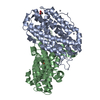
| ||||||||
|---|---|---|---|---|---|---|---|---|---|
| 1 |
| ||||||||
| Unit cell |
|
-Components
| #1: Protein | Mass: 43426.863 Da / Num. of mol.: 2 Source method: isolated from a genetically manipulated source Source: (gene. exp.) Escherichia coli (E. coli) / Plasmid: pTB2 / Production host: Escherichia coli (E. coli)References: UniProt: P69924, ribonucleoside-diphosphate reductase#2: Chemical | ChemComp-FE / Iron  Mass: 55.845 Da / Num. of mol.: 4 / Source method: obtained synthetically / Formula: Fe Mass: 55.845 Da / Num. of mol.: 4 / Source method: obtained synthetically / Formula: Fe#3: Chemical | ChemComp-HG / Mercury (element)  Mass: 200.590 Da / Num. of mol.: 11 / Source method: obtained synthetically / Formula: Hg Mass: 200.590 Da / Num. of mol.: 11 / Source method: obtained synthetically / Formula: Hg#4: Chemical | ChemComp-GOL / | Glycerol  Mass: 92.094 Da / Num. of mol.: 1 / Source method: obtained synthetically / Formula: C3H8O3 Mass: 92.094 Da / Num. of mol.: 1 / Source method: obtained synthetically / Formula: C3H8O3#5: Water | ChemComp-HOH / | Water Mass: 18.015 Da / Num. of mol.: 878 / Source method: isolated from a natural source / Formula: H2O Mass: 18.015 Da / Num. of mol.: 878 / Source method: isolated from a natural source / Formula: H2O |
|---|
-Experimental details
-Experiment
| Experiment | Method: X-RAY DIFFRACTION / Number of used crystals: 1 |
|---|
- Sample preparation
| Crystal | Density Matthews: 2.06 Å3/Da / Density % sol: 40.41 % | ||||||||||||||||||||||||||||||||||||
|---|---|---|---|---|---|---|---|---|---|---|---|---|---|---|---|---|---|---|---|---|---|---|---|---|---|---|---|---|---|---|---|---|---|---|---|---|---|
| Crystal grow | Temperature: 298 K / Method: vapor diffusion, hanging drop / pH: 6 Details: PEG 4000, MES, NaCl, EMTS, pH 6.0, VAPOR DIFFUSION, HANGING DROP, temperature 298K | ||||||||||||||||||||||||||||||||||||
| Crystal grow | *PLUS Temperature: 20 ℃ / Details: Nordlund, P. (1989) FEBS Lett., 258, 251. | ||||||||||||||||||||||||||||||||||||
| Components of the solutions | *PLUS
|
-Data collection
| Diffraction | Mean temperature: 100 K |
|---|---|
| Diffraction source | Source: SYNCHROTRON / Site: ESRF  / Beamline: BM1A / Wavelength: 0.87961 Å / Beamline: BM1A / Wavelength: 0.87961 Å |
| Detector | Type: MARRESEARCH / Detector: IMAGE PLATE / Date: Feb 8, 1999 |
| Radiation | Monochromator: SAGITALLY FOCUSED Si(111) / Protocol: SINGLE WAVELENGTH / Monochromatic (M) / Laue (L): M / Scattering type: x-ray |
| Radiation wavelength | Wavelength: 0.87961 Å / Relative weight: 1 |
| Reflection | Resolution: 1.42→40 Å / Num. all: 123371 / Num. obs: 123371 / % possible obs: 90.8 % / Observed criterion σ(F): 0 / Observed criterion σ(I): 0 / Biso Wilson estimate: 15.7 Å2 / Rsym value: 0.064 |
| Reflection shell | Resolution: 1.42→1.47 Å / Rmerge(I) obs: 0.288 / % possible all: 91.2 |
| Reflection | *PLUS Lowest resolution: 40 Å / Num. measured all: 603231 / Rmerge(I) obs: 0.064 |
| Reflection shell | *PLUS % possible obs: 91.2 % |
- Processing
| Software |
| ||||||||||||||||||||||||||||||||||||
|---|---|---|---|---|---|---|---|---|---|---|---|---|---|---|---|---|---|---|---|---|---|---|---|---|---|---|---|---|---|---|---|---|---|---|---|---|---|
| Refinement | Method to determine structure: MOLECULAR REPLACEMENT Starting model: reduced e. coli RNR R2 (1XIK) Resolution: 1.42→36.83 Å / Rfactor Rfree error: 0.002 / Isotropic thermal model: RESTRAINED / Cross valid method: THROUGHOUT / σ(F): 0 / Stereochemistry target values: Engh & Huber
| ||||||||||||||||||||||||||||||||||||
| Solvent computation | Solvent model: FLAT MODEL / Bsol: 69.6682 Å2 / ksol: 0.375864 e/Å3 | ||||||||||||||||||||||||||||||||||||
| Displacement parameters | Biso mean: 22.4 Å2
| ||||||||||||||||||||||||||||||||||||
| Refine analyze |
| ||||||||||||||||||||||||||||||||||||
| Refinement step | Cycle: LAST / Resolution: 1.42→36.83 Å
| ||||||||||||||||||||||||||||||||||||
| Refine LS restraints |
| ||||||||||||||||||||||||||||||||||||
| LS refinement shell | Resolution: 1.42→1.51 Å / Rfactor Rfree error: 0.007 / Total num. of bins used: 6
| ||||||||||||||||||||||||||||||||||||
| Xplor file |
| ||||||||||||||||||||||||||||||||||||
| Refinement | *PLUS Lowest resolution: 40 Å / % reflection Rfree: 5 % / Rfactor Rwork: 0.16 | ||||||||||||||||||||||||||||||||||||
| Solvent computation | *PLUS | ||||||||||||||||||||||||||||||||||||
| Displacement parameters | *PLUS | ||||||||||||||||||||||||||||||||||||
| Refine LS restraints | *PLUS
|
