 ムービー
ムービー コントローラー


+ データを開く

- 基本情報
基本情報
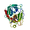
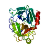
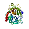
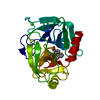
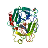
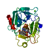
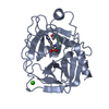
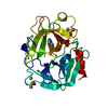
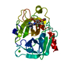
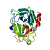
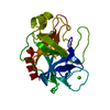
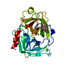
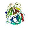
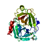
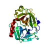
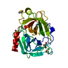
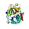
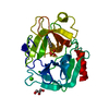
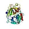
| 登録情報 | データベース: PDB / ID: 1mtu | ||||||
|---|---|---|---|---|---|---|---|
| タイトル | FACTOR XA SPECIFIC INHIBITOR IN COMPLEX WITH BOVINE TRYPSIN | ||||||
 要素 要素 | TRYPSIN トリプシン トリプシン | ||||||
 キーワード キーワード | SERINE PROTEASE (セリンプロテアーゼ) / HYDROLASE (加水分解酵素) | ||||||
| 機能・相同性 |  機能・相同性情報トリプシン / serpin family protein binding / serine protease inhibitor complex / 消化 / endopeptidase activity / serine-type endopeptidase activity / タンパク質分解 / extracellular space / metal ion binding 機能・相同性情報トリプシン / serpin family protein binding / serine protease inhibitor complex / 消化 / endopeptidase activity / serine-type endopeptidase activity / タンパク質分解 / extracellular space / metal ion binding類似検索 - 分子機能 | ||||||
| 生物種 |  Bos taurus (ウシ) Bos taurus (ウシ) | ||||||
| 手法 | X線回折 / DIFFERENCE FOURIER / 解像度: 1.9 Å | ||||||
 データ登録者 データ登録者 | Stubbs, M.T. | ||||||
 引用 引用 | ジャーナル: FEBS Lett. / 年: 1995 タイトル: Crystal structures of factor Xa specific inhibitors in complex with trypsin: structural grounds for inhibition of factor Xa and selectivity against thrombin. 著者: Stubbs, M.T. / Huber, R. / Bode, W. #1: ジャーナル: J.Mol.Biol. / 年: 1997タイトル: The Second Kunitz Domain of Human Tissue Factor Pathway Inhibitor. Cloning, Structure Determination and Interaction with Factor Xa 著者: Burgering, M.J. / Orbons, L.P. / Van Der Doelen, A. / Mulders, J. / Theunissen, H.J. / Grootenhuis, P.D. / Bode, W. / Huber, R. / Stubbs, M.T. #2: ジャーナル: Embo J. / 年: 1996タイトル: The Ornithodorin-Thrombin Crystal Structure, a Key to the Tap Enigma? 著者: Van De Locht, A. / Stubbs, M.T. / Bode, W. / Friedrich, T. / Bollschweiler, C. / Hoffken, W. / Huber, R. #3: ジャーナル: Curr.Pharm.Des. / 年: 1996タイトル: Structural Aspects of Factor Xa Inhibition 著者: Stubbs, M.T. #4: ジャーナル: J.Biol.Chem. / 年: 1996タイトル: X-Ray Structure of Active Site-Inhibited Clotting Factor Xa. Implications for Drug Design and Substrate Recognition 著者: Brandstetter, H. / Kuhne, A. / Bode, W. / Huber, R. / Von Der Saal, W. / Wirthensohn, K. / Engh, R.A. #5: ジャーナル: J.Mol.Biol. / 年: 1993タイトル: Structure of Human Des(1-45) Factor Xa at 2.2 A Resolution 著者: Padmanabhan, K. / Padmanabhan, K.P. / Tulinsky, A. / Park, C.H. / Bode, W. / Huber, R. / Blankenship, D.T. / Cardin, A.D. / Kisiel, W. #6: ジャーナル: FEBS Lett. / 年: 1991タイトル: Geometry of Binding of the N Alpha-Tosylated Piperidides of M-Amidino-, P-Amidino-and P-Guanidino Phenylalanine to Thrombin and Trypsin. X-Ray Crystal Structures of Their Trypsin ...タイトル: Geometry of Binding of the N Alpha-Tosylated Piperidides of M-Amidino-, P-Amidino-and P-Guanidino Phenylalanine to Thrombin and Trypsin. X-Ray Crystal Structures of Their Trypsin Complexes and Modeling of Their Thrombin Complexes 著者: Turk, D. / Sturzebecher, J. / Bode, W. #7: ジャーナル: Eur.J.Biochem. / 年: 1990タイトル: Geometry of Binding of the Benzamidine-and Arginine-Based Inhibitors N Alpha-(2-Naphthyl-Sulphonyl-Glycyl)-Dl-P-Amidinophenylalanyl-Piperidine (Napap) and (2R,4R)-4-Methyl-1-[N Alpha-(3- ...タイトル: Geometry of Binding of the Benzamidine-and Arginine-Based Inhibitors N Alpha-(2-Naphthyl-Sulphonyl-Glycyl)-Dl-P-Amidinophenylalanyl-Piperidine (Napap) and (2R,4R)-4-Methyl-1-[N Alpha-(3-Methyl-1,2,3,4-Tetrahydro-8-Quinolinesulphonyl)-L-Arginyl]-2-Piperidine to Carboxylic Acid (Mqpa) Human Alpha-Thrombin. X-Ray Crystallographic Determination of the Napap-Trypsin Complex and Modeling of Napap-Thrombin and Mqpa-Thrombin 著者: Bode, W. / Turk, D. / Sturzebecher, J. #8: ジャーナル: J.Mol.Biol. / 年: 1989タイトル: Crystal Structure of Bovine Beta-Trypsin at 1.5 A Resolution in a Crystal Form with Low Molecular Packing Density. Active Site Geometry, Ion Pairs and Solvent Structure 著者: Bartunik, H.D. / Summers, L.J. / Bartsch, H.H. #9: ジャーナル: J.Mol.Biol. / 年: 1975タイトル: The Refined Crystal Structure of Bovine Beta-Trypsin at 1.8 A Resolution. II. Crystallographic Refinement, Calcium Binding Site, Benzamidine Binding Site and Active Site at Ph 7.0 著者: Bode, W. / Schwager, P. | ||||||
| 履歴 |
|
- 構造の表示
構造の表示
| 構造ビューア | 分子: MolmilJmol/JSmol |
|---|
- ダウンロードとリンク
-ダウンロード
| PDBx/mmCIF形式 | 1mtu.cif.gz | 57.5 KB | 表示 | PDBx/mmCIF形式 |
|---|---|---|---|---|
| PDB形式 | pdb1mtu.ent.gz | 44.4 KB | 表示 | PDB形式 |
| PDBx/mmJSON形式 | 1mtu.json.gz | ツリー表示 | PDBx/mmJSON形式 | |
| その他 |  その他のダウンロード その他のダウンロード |
-検証レポート
| アーカイブディレクトリ | https://data.pdbj.org/pub/pdb/validation_reports/mt/1mtuftp://data.pdbj.org/pub/pdb/validation_reports/mt/1mtu | HTTPS FTP |
|---|
-関連構造データ


-リンク
 PDBj
PDBj

- 集合体
集合体
| 登録構造単位 | 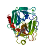
| ||||||||
|---|---|---|---|---|---|---|---|---|---|
| 1 |
| ||||||||
| 単位格子 |
|
-要素
| #1: タンパク質 | トリプシン 分子量: 23324.287 Da / 分子数: 1 / 由来タイプ: 天然 / 由来: (天然) Bos taurus (ウシ) / 参照: UniProt: P00760, トリプシン |
|---|---|
| #2: 化合物 | ChemComp-CA /   分子量: 40.078 Da / 分子数: 1 / 由来タイプ: 合成 / 式: Ca 分子量: 40.078 Da / 分子数: 1 / 由来タイプ: 合成 / 式: Ca |
| #3: 化合物 | ChemComp-BX3 / (+)-  分子量: 458.552 Da / 分子数: 1 / 由来タイプ: 合成 / 式: C27H30N4O3 分子量: 458.552 Da / 分子数: 1 / 由来タイプ: 合成 / 式: C27H30N4O3 |
| #4: 水 | ChemComp-HOH / 水 分子量: 18.015 Da / 分子数: 162 / 由来タイプ: 天然 / 式: H2O 分子量: 18.015 Da / 分子数: 162 / 由来タイプ: 天然 / 式: H2O |
-実験情報
-実験
| 実験 | 手法: X線回折 / 使用した結晶の数: 1 |
|---|
- 試料調製
試料調製
| 結晶 | マシュー密度: 3 Å3/Da / 溶媒含有率: 58.98 % | ||||||||||||||||||||||||||||||
|---|---|---|---|---|---|---|---|---|---|---|---|---|---|---|---|---|---|---|---|---|---|---|---|---|---|---|---|---|---|---|---|
| 結晶化 | pH: 6 / 詳細: 1.8M AMMONIUM SULFATE, PH 6.0 | ||||||||||||||||||||||||||||||
| 結晶化 | *PLUS 温度: 20 ℃ / 手法: 蒸気拡散法 / 詳細: Bode, W., (1990) Eur.J.Biochem., 193, 175. | ||||||||||||||||||||||||||||||
| 溶液の組成 | *PLUS
|
-データ収集
| 回折 | 平均測定温度: 287 K |
|---|---|
| 放射光源 | 由来: 回転陽極 / タイプ: RIGAKU / 波長: 1.5418 |
| 検出器 | タイプ: SIEMENS / 検出器: AREA DETECTOR / 日付: 1994年11月1日 / 詳細: MIRRORS |
| 放射 | モノクロメーター: NI FILTER / 単色(M)・ラウエ(L): M / 散乱光タイプ: x-ray |
| 放射波長 | 波長: 1.5418 Å / 相対比: 1 |
| 反射 | 解像度: 1.9→20 Å / Num. obs: 22069 / % possible obs: 96.8 % / Observed criterion σ(I): 2 / 冗長度: 2.2 % / Rmerge(I) obs: 0.029 / Rsym value: 0.029 / Net I/σ(I): 12 |
| 反射 シェル | 解像度: 1.9→2 Å / 冗長度: 1.7 % / Rmerge(I) obs: 0.087 / Mean I/σ(I) obs: 3 / Rsym value: 0.087 / % possible all: 92.6 |
| 反射 | *PLUS Num. measured all: 49006 |
- 解析
解析
| ソフトウェア |
| ||||||||||||||||||||||||||||||||||||||||||||||||||||||||||||
|---|---|---|---|---|---|---|---|---|---|---|---|---|---|---|---|---|---|---|---|---|---|---|---|---|---|---|---|---|---|---|---|---|---|---|---|---|---|---|---|---|---|---|---|---|---|---|---|---|---|---|---|---|---|---|---|---|---|---|---|---|---|
| 精密化 | 構造決定の手法: DIFFERENCE FOURIER / 解像度: 1.9→8 Å / Data cutoff high absF: 10000000 / Data cutoff low absF: 0.001 / σ(F): 3 /
| ||||||||||||||||||||||||||||||||||||||||||||||||||||||||||||
| Refine analyze | Luzzati d res low obs: 8 Å | ||||||||||||||||||||||||||||||||||||||||||||||||||||||||||||
| 精密化ステップ | サイクル: LAST / 解像度: 1.9→8 Å
| ||||||||||||||||||||||||||||||||||||||||||||||||||||||||||||
| 拘束条件 |
| ||||||||||||||||||||||||||||||||||||||||||||||||||||||||||||
| LS精密化 シェル | 解像度: 1.9→1.93 Å / Total num. of bins used: 20 /
| ||||||||||||||||||||||||||||||||||||||||||||||||||||||||||||
| Xplor file |
| ||||||||||||||||||||||||||||||||||||||||||||||||||||||||||||
| ソフトウェア | *PLUS 名称: X-PLOR / バージョン: 3.1 / 分類: refinement | ||||||||||||||||||||||||||||||||||||||||||||||||||||||||||||
| 拘束条件 | *PLUS
|
