 Movie
Movie Controller
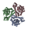


+ Open data

- Basic information
| Entry | Database: PDB / ID: 1mpr | ||||||
|---|---|---|---|---|---|---|---|
| Title | MALTOPORIN FROM SALMONELLA TYPHIMURIUM | ||||||
 Components Components | MALTOPORIN | ||||||
 Keywords Keywords | MEMBRANE PROTEIN / SUGAR TRANSPORT / SPECIFIC PORIN / BETA BARREL | ||||||
| Function / homology |  Function and homology information Function and homology informationmaltodextrin transmembrane transporter activity / maltose transporting porin activity / polysaccharide transport / porin activity / pore complex / carbohydrate transmembrane transporter activity / monoatomic ion transport / cell outer membraneSimilarity search - Function | ||||||
| Biological species |  Salmonella typhimurium (bacteria) Salmonella typhimurium (bacteria) | ||||||
| Method | X-RAY DIFFRACTION / SYNCHROTRON / MOLECULAR REPLACEMENT / Resolution: 2.8 Å | ||||||
 Authors Authors | Meyer, J.E.W. / Schulz, G.E. | ||||||
 Citation Citation | Journal: J.Mol.Biol. / Year: 1997 Title: Structure of maltoporin from Salmonella typhimurium ligated with a nitrophenyl-maltotrioside. Authors: Meyer, J.E. / Hofnung, M. / Schulz, G.E. | ||||||
| History |
|
- Structure visualization
| Structure viewer | Molecule: MolmilJmol/JSmol |
|---|
- Downloads & links
-Download
| PDBx/mmCIF format | 1mpr.cif.gz | 260.6 KB | Display | PDBx/mmCIF format |
|---|---|---|---|---|
| PDB format | pdb1mpr.ent.gz | 213.3 KB | Display | PDB format |
| PDBx/mmJSON format | 1mpr.json.gz | Tree view | PDBx/mmJSON format | |
| Others |  Other downloads Other downloads |
-Validation report
| Arichive directory | https://data.pdbj.org/pub/pdb/validation_reports/mp/1mprftp://data.pdbj.org/pub/pdb/validation_reports/mp/1mpr | HTTPS FTP |
|---|
-Related structure data
| Related structure data |  2mprC  1malS S: Starting model for refinement C: citing same article ( |
|---|---|
| Similar structure data |


-Links
 PDBj
PDBj- Assembly
| Deposited unit | 
| ||||||||||||
|---|---|---|---|---|---|---|---|---|---|---|---|---|---|
| 1 |
| ||||||||||||
| Unit cell |
| ||||||||||||
| Noncrystallographic symmetry (NCS) | NCS oper:
| ||||||||||||
| Details | THE FOLLOWING TRANSFORMATION WILL TRANSFORM THE WATERS OF CHAIN A INTO THE WATERS OF CHAIN B. MTRIX1 -0.485848 -0.865005 -0.125370 65.39400 MTRIX2 0.864287 -0.496828 0.078542 99.80500 MTRIX3 -0.130227 -0.070196 0.988996 5.42100 THE FOLLOWING TRANSFORMATION WILL TRANSFORM THE WATERS OF CHAIN A INTO THE WATERS OF CHAIN C. MTRIX1 -0.488651 0.862290 -0.132950 -53.38000 MTRIX2 -0.862650 -0.500312 -0.074311 107.08700 MTRIX3 -0.130594 0.078377 0.988333 -4.78000 |
-Components
| #1: Protein | / LAM-B / MAL-L Mass: 48064.535 Da / Num. of mol.: 3 / Source method: isolated from a natural source / Source: (natural) Salmonella typhimurium (bacteria) / Cellular location: OUTER MEMBRANE / Strain: SL3749 / References: UniProt: P26466#2: Chemical | ChemComp-CA / |   Mass: 40.078 Da / Num. of mol.: 1 / Source method: obtained synthetically / Formula: Ca Mass: 40.078 Da / Num. of mol.: 1 / Source method: obtained synthetically / Formula: Ca#3: Water | ChemComp-HOH / | Water Mass: 18.015 Da / Num. of mol.: 456 / Source method: isolated from a natural source / Formula: H2O Mass: 18.015 Da / Num. of mol.: 456 / Source method: isolated from a natural source / Formula: H2O |
|---|
-Experimental details
-Experiment
| Experiment | Method: X-RAY DIFFRACTION / Number of used crystals: 1 |
|---|
- Sample preparation
| Crystal | Density Matthews: 4.3 Å3/Da / Density % sol: 71 % | ||||||||||||||||||||||||||||||||||||||||||||||||||||||
|---|---|---|---|---|---|---|---|---|---|---|---|---|---|---|---|---|---|---|---|---|---|---|---|---|---|---|---|---|---|---|---|---|---|---|---|---|---|---|---|---|---|---|---|---|---|---|---|---|---|---|---|---|---|---|---|
| Crystal grow | Method: vapor diffusion, hanging drop Details: MALTOPORIN WAS CRYSTALLIZED BY HANGING-DROP METHOD. DROP: 5-8 MG/ML PROTEIN, 0.3% C8E4, 0.8% C6DAO, 1 MM CACL2, 1MM MGCL2, 14-18% PEG 1500, 0.02% NAN3 RESERVOIR: 28-32% PEG 1500, vapor diffusion - hanging drop | ||||||||||||||||||||||||||||||||||||||||||||||||||||||
| Crystal grow | *PLUS Temperature: 18 ℃ / Method: vapor diffusion, hanging drop | ||||||||||||||||||||||||||||||||||||||||||||||||||||||
| Components of the solutions | *PLUS
|
-Data collection
| Diffraction | Mean temperature: 298 K |
|---|---|
| Diffraction source | Source: SYNCHROTRON / Site: EMBL/DESY, HAMBURG  / Beamline: X31 / Wavelength: 1 / Beamline: X31 / Wavelength: 1 |
| Detector | Type: MARRESEARCH / Detector: IMAGE PLATE / Date: May 14, 1994 |
| Radiation | Monochromatic (M) / Laue (L): M / Scattering type: x-ray |
| Radiation wavelength | Wavelength: 1 Å / Relative weight: 1 |
| Reflection | Resolution: 2.8→20 Å / Num. obs: 59257 / % possible obs: 95 % / Redundancy: 2.9 % / Biso Wilson estimate: 45 Å2 / Rsym value: 0.072 / Net I/σ(I): 7.1 |
| Reflection shell | Resolution: 2.77→2.84 Å / Redundancy: 2.4 % / Mean I/σ(I) obs: 3.5 / Rsym value: 0.259 / % possible all: 91.4 |
| Reflection | *PLUS Num. measured all: 171366 / Rmerge(I) obs: 0.072 |
| Reflection shell | *PLUS % possible obs: 91.4 % / Rmerge(I) obs: 0.259 |
- Processing
| Software |
| ||||||||||||||||||||||||||||||||||||||||||||||||||||||||||||
|---|---|---|---|---|---|---|---|---|---|---|---|---|---|---|---|---|---|---|---|---|---|---|---|---|---|---|---|---|---|---|---|---|---|---|---|---|---|---|---|---|---|---|---|---|---|---|---|---|---|---|---|---|---|---|---|---|---|---|---|---|---|
| Refinement | Method to determine structure: MOLECULAR REPLACEMENT Starting model: PDB ENTRY 1MAL Resolution: 2.8→10 Å / Rfactor Rfree error: 0.0028 / Data cutoff high absF: 10000000 / Data cutoff low absF: 0 / Isotropic thermal model: RESTRAINED Cross valid method: THROUGHOUT EXCEPT FOR VERY LAST ROUND (REFINEMENT AGAINST ALL REFLECTIONS) σ(F): 0 Details: ALL SIDE CHAIN ATOMS WITHOUT DENSITY WERE ASSIGNED ZERO OCCUPANCY. B-VALUES OF WILSON PLOT WAS CALCULATED WITH THE CCP4 PROGRAM WILSON.
| ||||||||||||||||||||||||||||||||||||||||||||||||||||||||||||
| Displacement parameters | Biso mean: 21.1 Å2 | ||||||||||||||||||||||||||||||||||||||||||||||||||||||||||||
| Refine analyze | Luzzati d res low obs: 3.56 Å / Luzzati sigma a obs: 0.38 Å | ||||||||||||||||||||||||||||||||||||||||||||||||||||||||||||
| Refinement step | Cycle: LAST / Resolution: 2.8→10 Å
| ||||||||||||||||||||||||||||||||||||||||||||||||||||||||||||
| Refine LS restraints |
| ||||||||||||||||||||||||||||||||||||||||||||||||||||||||||||
| Refine LS restraints NCS | NCS model details: RESTRAINTS / Rms dev Biso : 2 Å2 / Rms dev position: 0.2 Å / Weight Biso : 0.2 / Weight position: 100 | ||||||||||||||||||||||||||||||||||||||||||||||||||||||||||||
| LS refinement shell | Resolution: 2.8→2.9 Å / Rfactor Rfree error: 0.012 / Total num. of bins used: 10
| ||||||||||||||||||||||||||||||||||||||||||||||||||||||||||||
| Xplor file |
| ||||||||||||||||||||||||||||||||||||||||||||||||||||||||||||
| Software | *PLUS Name: X-PLOR / Classification: refinement | ||||||||||||||||||||||||||||||||||||||||||||||||||||||||||||
| Refinement | *PLUS | ||||||||||||||||||||||||||||||||||||||||||||||||||||||||||||
| Solvent computation | *PLUS | ||||||||||||||||||||||||||||||||||||||||||||||||||||||||||||
| Displacement parameters | *PLUS | ||||||||||||||||||||||||||||||||||||||||||||||||||||||||||||
| Refine LS restraints | *PLUS
|
