 Movie
Movie Controller

[English] 日本語
 Yorodumi
- PDB-1mmm: DISTINCT METAL ENVIRONMENT IN IRON-SUBSTITUTED MANGANESE SUPEROXI... -
+ Open data

- Basic information
| Entry | Database: PDB / ID: 1mmm | ||||||
|---|---|---|---|---|---|---|---|
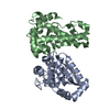
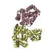
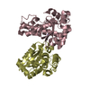
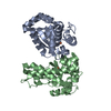
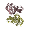
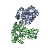
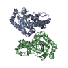
| Title | DISTINCT METAL ENVIRONMENT IN IRON-SUBSTITUTED MANGANESE SUPEROXIDE DISMUTASE PROVIDES A STRUCTURAL BASIS OF METAL SPECIFICITY | ||||||
 Components Components | PROTEIN (IRON-SUBSTITUTED MANGANESE SUPEROXIDE DISMUTASE) | ||||||
 Keywords Keywords |  OXIDOREDUCTASE / IRON-SUBSTITUTED MANGANESE SUPEROXIDE DISMUTASE OXIDOREDUCTASE / IRON-SUBSTITUTED MANGANESE SUPEROXIDE DISMUTASE | ||||||
| Function / homology |  Function and homology information Function and homology informationcellular response to selenium ion / response to acidic pH / superoxide metabolic process / superoxide dismutase / superoxide dismutase activity / antioxidant activity / removal of superoxide radicals / manganese ion binding / response to heat / response to oxidative stress ...cellular response to selenium ion / response to acidic pH / superoxide metabolic process / superoxide dismutase / superoxide dismutase activity / antioxidant activity / removal of superoxide radicals / manganese ion binding / response to heat / response to oxidative stress / protein homodimerization activity / DNA binding / metal ion binding / cytosol / cytoplasmSimilarity search - Function | ||||||
| Biological species |  Escherichia coli (E. coli) Escherichia coli (E. coli) | ||||||
| Method | X-RAY DIFFRACTION / MOLECULAR REPLACEMENT / Resolution: 2.2 Å | ||||||
 Authors Authors | Edwards, R.A. / Whittaker, M.M. / Whittaker, J.W. / Jameson, G.B. / Baker, E.N. | ||||||
 Citation Citation | Journal: J.Am.Chem.Soc. / Year: 1998 Title: Distinct Metal Environment in Fe-Substituted Manganese Superoxide Dismutase Provides a Structural Basis of Metal Specificity Authors: Edwards, R.A. / Whittaker, M.M. / Whittaker, J.W. / Jameson, G.B. / Baker, E.N. | ||||||
| History |
|
- Structure visualization
| Structure viewer | Molecule: MolmilJmol/JSmol |
|---|
- Downloads & links
-Download
| PDBx/mmCIF format | 1mmm.cif.gz | 97 KB | Display | PDBx/mmCIF format |
|---|---|---|---|---|
| PDB format | pdb1mmm.ent.gz | 74.1 KB | Display | PDB format |
| PDBx/mmJSON format | 1mmm.json.gz | Tree view | PDBx/mmJSON format | |
| Others |  Other downloads Other downloads |
-Validation report
| Arichive directory | https://data.pdbj.org/pub/pdb/validation_reports/mm/1mmmftp://data.pdbj.org/pub/pdb/validation_reports/mm/1mmm | HTTPS FTP |
|---|
-Related structure data
| Related structure data | 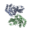 1vewS S: Starting model for refinement |
|---|---|
| Similar structure data |


-Links
 PDBj
PDBj


- Assembly
| Deposited unit | 
| ||||||||
|---|---|---|---|---|---|---|---|---|---|
| 1 |
| ||||||||
| Unit cell |
| ||||||||
| Noncrystallographic symmetry (NCS) | NCS oper: (Code: given Matrix: (0.729903, 0.132591, 0.670567), Vector : |
-Components
| #1: Protein | Mass: 22996.877 Da / Num. of mol.: 2 / Source method: isolated from a natural source / Source: (natural) Escherichia coli (E. coli) / Strain: AB2463/PDT1-5 / References: UniProt: P00448, superoxide dismutase#2: Chemical | Hydroxide  Mass: 17.007 Da / Num. of mol.: 3 / Source method: obtained synthetically / Formula: HO Mass: 17.007 Da / Num. of mol.: 3 / Source method: obtained synthetically / Formula: HO#3: Chemical | Iron  Mass: 55.845 Da / Num. of mol.: 2 / Source method: obtained synthetically / Formula: Fe Mass: 55.845 Da / Num. of mol.: 2 / Source method: obtained synthetically / Formula: Fe#4: Water | ChemComp-HOH / | Water Mass: 18.015 Da / Num. of mol.: 284 / Source method: isolated from a natural source / Formula: H2O Mass: 18.015 Da / Num. of mol.: 284 / Source method: isolated from a natural source / Formula: H2O |
|---|
-Experimental details
-Experiment
| Experiment | Method: X-RAY DIFFRACTION / Number of used crystals: 1 |
|---|
- Sample preparation
| Crystal | Density Matthews: 2.31 Å3/Da / Density % sol: 46.73 % | ||||||||||||||||||||||||||||||
|---|---|---|---|---|---|---|---|---|---|---|---|---|---|---|---|---|---|---|---|---|---|---|---|---|---|---|---|---|---|---|---|
| Crystal grow | pH: 8.75 / Details: pH 8.75 | ||||||||||||||||||||||||||||||
| Crystal grow | *PLUS pH: 8.5 / Method: vapor diffusion, hanging dropDetails: Edwards, R.A., (1998) J. Biol. Inorg. Biol., 3, 161. | ||||||||||||||||||||||||||||||
| Components of the solutions | *PLUS
|
-Data collection
| Diffraction | Mean temperature: 110 K |
|---|---|
| Diffraction source | Source: ROTATING ANODE / Type: RIGAKU RU200 / Wavelength: 1.5418 |
| Detector | Type: RIGAKU RAXIS IIC / Detector: IMAGE PLATE / Date: Jan 15, 1998 |
| Radiation | Monochromator: GRAPHITE / Protocol: SINGLE WAVELENGTH / Monochromatic (M) / Laue (L): M / Scattering type: x-ray |
| Radiation wavelength | Wavelength: 1.5418 Å / Relative weight: 1 |
| Reflection | Resolution: 2.2→100 Å / Num. obs: 20726 / % possible obs: 93.2 % / Redundancy: 4.2 % / Rmerge(I) obs: 0.09 / Rsym value: 0.09 / Net I/σ(I): 14 |
| Reflection shell | Resolution: 2.2→2.3 Å / Rmerge(I) obs: 0.266 / Mean I/σ(I) obs: 4.6 / Rsym value: 0.266 / % possible all: 82.6 |
| Reflection | *PLUS Rmerge(I) obs: 0.09 |
| Reflection shell | *PLUS % possible obs: 82.6 % |
- Processing
| Software |
| ||||||||||||||||||||||||||||||||||||||||||||||||||||||||||||
|---|---|---|---|---|---|---|---|---|---|---|---|---|---|---|---|---|---|---|---|---|---|---|---|---|---|---|---|---|---|---|---|---|---|---|---|---|---|---|---|---|---|---|---|---|---|---|---|---|---|---|---|---|---|---|---|---|---|---|---|---|---|
| Refinement | Method to determine structure: MOLECULAR REPLACEMENT Starting model: 1VEW Resolution: 2.2→100 Å / Cross valid method: THROUGHOUT / σ(F): 0
| ||||||||||||||||||||||||||||||||||||||||||||||||||||||||||||
| Displacement parameters |
| ||||||||||||||||||||||||||||||||||||||||||||||||||||||||||||
| Refinement step | Cycle: LAST / Resolution: 2.2→100 Å
| ||||||||||||||||||||||||||||||||||||||||||||||||||||||||||||
| Refine LS restraints |
| ||||||||||||||||||||||||||||||||||||||||||||||||||||||||||||
| Refine LS restraints NCS | NCS model details: RESTRAINTS | ||||||||||||||||||||||||||||||||||||||||||||||||||||||||||||
| Software | *PLUS Name: CNS / Version: 0.3C / Classification: refinement | ||||||||||||||||||||||||||||||||||||||||||||||||||||||||||||
| Refinement | *PLUS Highest resolution: 2.2 Å / Lowest resolution: 100 Å / σ(F): 0 / % reflection Rfree: 7 % | ||||||||||||||||||||||||||||||||||||||||||||||||||||||||||||
| Solvent computation | *PLUS | ||||||||||||||||||||||||||||||||||||||||||||||||||||||||||||
| Displacement parameters | *PLUS | ||||||||||||||||||||||||||||||||||||||||||||||||||||||||||||
| Refine LS restraints | *PLUS Type: c_angle_deg / Dev ideal: 1.3 |
