 Movie
Movie Controller

[English] 日本語
 Yorodumi
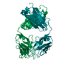
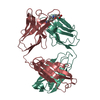
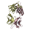
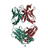
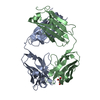
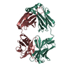
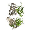
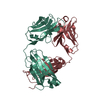
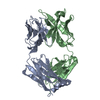
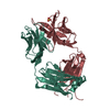
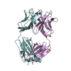
- PDB-1mlb: MONOCLONAL ANTIBODY FAB D44.1 RAISED AGAINST CHICKEN EGG-WHITE LY... -
+ Open data

- Basic information
| Entry | Database: PDB / ID: 1mlb | ||||||
|---|---|---|---|---|---|---|---|
| Title | MONOCLONAL ANTIBODY FAB D44.1 RAISED AGAINST CHICKEN EGG-WHITE LYSOZYME | ||||||
 Components Components |
| ||||||
 Keywords Keywords |  IMMUNE SYSTEM / IMMUNOGLOBULIN IMMUNE SYSTEM / IMMUNOGLOBULIN | ||||||
| Function / homology |  Function and homology information Function and homology information | ||||||
| Biological species |  Mus musculus (house mouse) Mus musculus (house mouse) | ||||||
| Method | X-RAY DIFFRACTION / Resolution: 2.1 Å | ||||||
 Authors Authors | Braden, B.C. / Souchon, H. / Eisele, J.-L. / Bentley, G.A. / Bhat, T.N. / Navaza, J. / Poljak, R.J. | ||||||
 Citation Citation | Journal: J.Mol.Biol. / Year: 1994 Title: Three-dimensional structures of the free and the antigen-complexed Fab from monoclonal anti-lysozyme antibody D44.1. Authors: Braden, B.C. / Souchon, H. / Eisele, J.L. / Bentley, G.A. / Bhat, T.N. / Navaza, J. / Poljak, R.J. #1: Journal: J.Mol.Biol. / Year: 1988Title: Crystallization and Preliminary X-Ray Diffraction Studies of Two Antigen-Antibody (Lysozyme-Fab) Complexes Authors: Fischmann, T. / Souchon, H. / Riottot, M.-M. / Tello, D. / Poljak, R.J. | ||||||
| History |
|
- Structure visualization
| Structure viewer | Molecule: MolmilJmol/JSmol |
|---|
- Downloads & links
-Download
| PDBx/mmCIF format | 1mlb.cif.gz | 94.2 KB | Display | PDBx/mmCIF format |
|---|---|---|---|---|
| PDB format | pdb1mlb.ent.gz | 75.6 KB | Display | PDB format |
| PDBx/mmJSON format | 1mlb.json.gz | Tree view | PDBx/mmJSON format | |
| Others |  Other downloads Other downloads |
-Validation report
| Arichive directory | https://data.pdbj.org/pub/pdb/validation_reports/ml/1mlbftp://data.pdbj.org/pub/pdb/validation_reports/ml/1mlb | HTTPS FTP |
|---|
-Related structure data


-Links
 PDBj
PDBj


- Assembly
| Deposited unit | 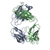
| ||||||||
|---|---|---|---|---|---|---|---|---|---|
| 1 |
| ||||||||
| Unit cell |
| ||||||||
| Atom site foot note | 1: CIS PROLINE - PRO A 8 / 2: CIS PROLINE - PRO A 95 / 3: CIS PROLINE - PRO A 141 / 4: CIS PROLINE - PRO B 150 / 5: CIS PROLINE - PRO B 152 / 6: CIS PROLINE - PRO B 192 |
-Components
| #1: Antibody | Mass: 23609.846 Da / Num. of mol.: 1 Source method: isolated from a genetically manipulated source Source: (gene. exp.) Mus musculus (house mouse) / References: UniProt: P01837 |
|---|---|
| #2: Antibody | Mass: 23215.848 Da / Num. of mol.: 1 Source method: isolated from a genetically manipulated source Source: (gene. exp.) Mus musculus (house mouse) / References: PIR: PC4202 |
| #3: Water | ChemComp-HOH / Water Mass: 18.015 Da / Num. of mol.: 115 / Source method: isolated from a natural source / Formula: H2O Mass: 18.015 Da / Num. of mol.: 115 / Source method: isolated from a natural source / Formula: H2O |
| Compound details | VL RESIDUE SER 30 IS IN THE SECOND POSITION OF A II'-TYPE TURN. VL RESIDUE VAL 51 IS THE SECOND ...VL RESIDUE SER 30 IS IN THE SECOND POSITION OF A II'-TYPE TURN. VL RESIDUE VAL 51 IS THE SECOND RESIDUE (I+1) OF A MODIFIED GAMMA TURN (CLASS 3). |
-Experimental details
-Experiment
| Experiment | Method: X-RAY DIFFRACTION / Number of used crystals: 1 |
|---|
- Sample preparation
| Crystal | Density Matthews: 2.49 Å3/Da / Density % sol: 50.62 % | ||||||||||||||||||||
|---|---|---|---|---|---|---|---|---|---|---|---|---|---|---|---|---|---|---|---|---|---|
| Crystal grow | *PLUS pH: 6 / Method: vapor diffusion, hanging drop | ||||||||||||||||||||
| Components of the solutions | *PLUS
|
-Data collection
| Diffraction source | Wavelength: 1.5418 Å |
|---|---|
| Detector | Type: SIEMENS / Detector: AREA DETECTOR |
| Radiation | Scattering type: x-ray |
| Radiation wavelength | Wavelength: 1.5418 Å / Relative weight: 1 |
| Reflection | Num. obs: 26986 / % possible obs: 70 % / Redundancy: 4 % / Rmerge(I) obs: 0.1 |
| Reflection | *PLUS Highest resolution: 2.1 Å / Rmerge(I) obs: 0.1 / Num. measured all: 106016 |
- Processing
| Software |
| ||||||||||||||||||||||||||||||||||||||||||||||||||||||||||||||||||||||||||||||||||||
|---|---|---|---|---|---|---|---|---|---|---|---|---|---|---|---|---|---|---|---|---|---|---|---|---|---|---|---|---|---|---|---|---|---|---|---|---|---|---|---|---|---|---|---|---|---|---|---|---|---|---|---|---|---|---|---|---|---|---|---|---|---|---|---|---|---|---|---|---|---|---|---|---|---|---|---|---|---|---|---|---|---|---|---|---|---|
| Refinement | Resolution: 2.1→7 Å / σ(F): 2 Details: D44.1 IS CRYSTALLIZED AS THE FAB. CHAIN A INCLUDES THE VL AND CL DOMAINS. CHAIN B INCLUDES THE VH AND CH1 DOMAINS. CHAIN B RESIDUES 131 - 136, ASP 217 AND THE C-TERMINAL CYS 218 (ALL IN THE ...Details: D44.1 IS CRYSTALLIZED AS THE FAB. CHAIN A INCLUDES THE VL AND CL DOMAINS. CHAIN B INCLUDES THE VH AND CH1 DOMAINS. CHAIN B RESIDUES 131 - 136, ASP 217 AND THE C-TERMINAL CYS 218 (ALL IN THE CH1 DOMAIN) HAVE NO ELECTRON DENSITY AND, THEREFORE, THE ATOMIC POSITIONS FOR THESE RESIDUES SHOULD BE CONSIDERED ARBITRARY.
| ||||||||||||||||||||||||||||||||||||||||||||||||||||||||||||||||||||||||||||||||||||
| Displacement parameters | Biso mean: 27.1 Å2 | ||||||||||||||||||||||||||||||||||||||||||||||||||||||||||||||||||||||||||||||||||||
| Refinement step | Cycle: LAST / Resolution: 2.1→7 Å
| ||||||||||||||||||||||||||||||||||||||||||||||||||||||||||||||||||||||||||||||||||||
| Refine LS restraints |
|
