 Movie
Movie Controller


+ Open data

- Basic information
| Entry | Database: PDB / ID: 1mkj | ||||||
|---|---|---|---|---|---|---|---|
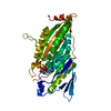
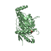
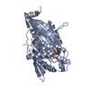
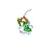
| Title | Human Kinesin Motor Domain With Docked Neck Linker | ||||||
 Components Components | KINESIN HEAVY CHAIN | ||||||
 Keywords Keywords | TRANSPORT PROTEIN / NECK LINKER / SWITCH II / MOTOR PROTEIN / MYOSIN / RELAY HELIX / MICROTUBULE / ATPASE | ||||||
| Function / homology |  Function and homology information Function and homology informationcytoplasm organization / cytolytic granule membrane / plus-end-directed vesicle transport along microtubule / anterograde neuronal dense core vesicle transport / anterograde dendritic transport of neurotransmitter receptor complex / mitocytosis / retrograde neuronal dense core vesicle transport / anterograde axonal protein transport / vesicle transport along microtubule / lysosome localization ...cytoplasm organization / cytolytic granule membrane / plus-end-directed vesicle transport along microtubule / anterograde neuronal dense core vesicle transport / anterograde dendritic transport of neurotransmitter receptor complex / mitocytosis / retrograde neuronal dense core vesicle transport / anterograde axonal protein transport / vesicle transport along microtubule / lysosome localization / positive regulation of potassium ion transport / natural killer cell mediated cytotoxicity / Kinesins / plus-end-directed microtubule motor activity / RHO GTPases activate KTN1 / stress granule disassembly / COPI-dependent Golgi-to-ER retrograde traffic / mitochondrion transport along microtubule / ciliary rootlet / centrosome localization / kinesin complex / synaptic vesicle transport / microtubule motor activity / microtubule-based movement / Insulin processing / centriolar satellite / Signaling by ALK fusions and activated point mutants / Nuclear events stimulated by ALK signaling in cancer / phagocytic vesicle / axon cytoplasm / MHC class II antigen presentation / dendrite cytoplasm / regulation of membrane potential / axon guidance / positive regulation of synaptic transmission, GABAergic / positive regulation of protein localization to plasma membrane / cellular response to type II interferon / microtubule binding / vesicle / microtubule / cadherin binding / protein-containing complex binding / perinuclear region of cytoplasm / ATP hydrolysis activity / mitochondrion / ATP binding / membrane / identical protein binding / cytosolSimilarity search - Function | ||||||
| Biological species |  Homo sapiens (human) Homo sapiens (human) | ||||||
| Method | X-RAY DIFFRACTION / SYNCHROTRON / MOLECULAR REPLACEMENT / Resolution: 2.7 Å | ||||||
 Authors Authors | Sindelar, C.V. / Budny, M.J. / Rice, S. / Naber, N. / Fletterick, R. / Cooke, R. | ||||||
 Citation Citation | Journal: Nat.Struct.Biol. / Year: 2002 Title: Two conformations in the human kinesin power stroke defined by X-ray crystallography and EPR spectroscopy. Authors: Sindelar, C.V. / Budny, M.J. / Rice, S. / Naber, N. / Fletterick, R. / Cooke, R. | ||||||
| History |
|
- Structure visualization
| Structure viewer | Molecule: MolmilJmol/JSmol |
|---|
- Downloads & links
-Download
| PDBx/mmCIF format | 1mkj.cif.gz | 80.1 KB | Display | PDBx/mmCIF format |
|---|---|---|---|---|
| PDB format | pdb1mkj.ent.gz | 60 KB | Display | PDB format |
| PDBx/mmJSON format | 1mkj.json.gz | Tree view | PDBx/mmJSON format | |
| Others |  Other downloads Other downloads |
-Validation report
| Arichive directory | https://data.pdbj.org/pub/pdb/validation_reports/mk/1mkjftp://data.pdbj.org/pub/pdb/validation_reports/mk/1mkj | HTTPS FTP |
|---|
-Related structure data
| Related structure data |  2kinS S: Starting model for refinement |
|---|---|
| Similar structure data |


-Links
 PDBj
PDBj


- Assembly
| Deposited unit | 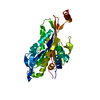
| ||||||||
|---|---|---|---|---|---|---|---|---|---|
| 1 |
| ||||||||
| Unit cell |
|
-Components
| #1: Protein | / ubiquitous kinesin heavy chain / UKHC Mass: 39318.406 Da / Num. of mol.: 1 / Fragment: Residues 1-349 Source method: isolated from a genetically manipulated source Source: (gene. exp.) Homo sapiens (human) / Plasmid: PET / Species (production host): Escherichia coli / Production host:  Escherichia coli BL21(DE3) (bacteria) / Strain (production host): BL21 (DE3) / References: UniProt: P33176 Escherichia coli BL21(DE3) (bacteria) / Strain (production host): BL21 (DE3) / References: UniProt: P33176 | ||||
|---|---|---|---|---|---|
| #2: Chemical | ChemComp-MG /   Mass: 24.305 Da / Num. of mol.: 1 / Source method: obtained synthetically / Formula: Mg Mass: 24.305 Da / Num. of mol.: 1 / Source method: obtained synthetically / Formula: Mg | ||||
| #3: Chemical | Sulfate  Mass: 96.063 Da / Num. of mol.: 2 / Source method: obtained synthetically / Formula: SO4 Mass: 96.063 Da / Num. of mol.: 2 / Source method: obtained synthetically / Formula: SO4#4: Chemical | ChemComp-ADP / | Adenosine diphosphate  Mass: 427.201 Da / Num. of mol.: 1 / Source method: obtained synthetically / Formula: C10H15N5O10P2 / Comment: ADP, energy-carrying molecule*YM Mass: 427.201 Da / Num. of mol.: 1 / Source method: obtained synthetically / Formula: C10H15N5O10P2 / Comment: ADP, energy-carrying molecule*YM#5: Water | ChemComp-HOH / | Water Mass: 18.015 Da / Num. of mol.: 7 / Source method: isolated from a natural source / Formula: H2O Mass: 18.015 Da / Num. of mol.: 7 / Source method: isolated from a natural source / Formula: H2O |
-Experimental details
-Experiment
| Experiment | Method: X-RAY DIFFRACTION / Number of used crystals: 1 |
|---|
- Sample preparation
| Crystal | Density Matthews: 3.18 Å3/Da / Density % sol: 61.34 % | |||||||||||||||||||||||||||||||||||||||||||||||||||||||||||||||
|---|---|---|---|---|---|---|---|---|---|---|---|---|---|---|---|---|---|---|---|---|---|---|---|---|---|---|---|---|---|---|---|---|---|---|---|---|---|---|---|---|---|---|---|---|---|---|---|---|---|---|---|---|---|---|---|---|---|---|---|---|---|---|---|---|
| Crystal grow | Temperature: 277 K / pH: 7.5 Details: 1.5-2 M Li2SO4, 25 mM HEPES pH 7.5, 0-50 mM KCl, pH 7.50, temperature 277K | |||||||||||||||||||||||||||||||||||||||||||||||||||||||||||||||
| Crystal grow | *PLUS pH: 6.8 / Method: vapor diffusion, hanging drop | |||||||||||||||||||||||||||||||||||||||||||||||||||||||||||||||
| Components of the solutions | *PLUS
|
-Data collection
| Diffraction | Mean temperature: 100 K |
|---|---|
| Diffraction source | Source: SYNCHROTRON / Site: ALS  / Beamline: 5.0.2 / Wavelength: 1.1 / Beamline: 5.0.2 / Wavelength: 1.1 |
| Detector | Type: ADSC QUANTUM 4 / Detector: CCD / Date: Oct 4, 2000 |
| Radiation | Protocol: SINGLE WAVELENGTH / Monochromatic (M) / Laue (L): M / Scattering type: x-ray |
| Radiation wavelength | Wavelength: 1.1 Å / Relative weight: 1 |
| Reflection | Resolution: 2.7→20 Å / Num. obs: 12579 / % possible obs: 99.8 % / Observed criterion σ(I): 0 / Biso Wilson estimate: 28.7 Å2 / Rsym value: 0.079 / Net I/σ(I): 23 |
| Reflection shell | Highest resolution: 2.7 Å / Mean I/σ(I) obs: 6.9 / Rsym value: 0.27 / % possible all: 100 |
| Reflection | *PLUS Lowest resolution: 20 Å / Num. obs: 14266 / Num. measured all: 247497 / Rmerge(I) obs: 0.079 |
| Reflection shell | *PLUS % possible obs: 100 % / Rmerge(I) obs: 0.27 |
- Processing
| Software |
| ||||||||||||||||||||||||||||||||||||||||||||||||||||||||||||||||||||||||||||||||
|---|---|---|---|---|---|---|---|---|---|---|---|---|---|---|---|---|---|---|---|---|---|---|---|---|---|---|---|---|---|---|---|---|---|---|---|---|---|---|---|---|---|---|---|---|---|---|---|---|---|---|---|---|---|---|---|---|---|---|---|---|---|---|---|---|---|---|---|---|---|---|---|---|---|---|---|---|---|---|---|---|---|
| Refinement | Method to determine structure: MOLECULAR REPLACEMENT Starting model: PDB ENTRY 2KIN Resolution: 2.7→20.03 Å / Rfactor Rfree error: 0.01 / Data cutoff high absF: 147240.76 / Data cutoff high rms absF: 147240.76 / Data cutoff low absF: 0 / Isotropic thermal model: RESTRAINED / Cross valid method: THROUGHOUT / σ(F): 0
| ||||||||||||||||||||||||||||||||||||||||||||||||||||||||||||||||||||||||||||||||
| Solvent computation | Solvent model: FLAT MODEL / Bsol: 23.5303 Å2 / ksol: 0.358848 e/Å3 | ||||||||||||||||||||||||||||||||||||||||||||||||||||||||||||||||||||||||||||||||
| Displacement parameters | Biso mean: 31.1 Å2
| ||||||||||||||||||||||||||||||||||||||||||||||||||||||||||||||||||||||||||||||||
| Refine analyze |
| ||||||||||||||||||||||||||||||||||||||||||||||||||||||||||||||||||||||||||||||||
| Refinement step | Cycle: LAST / Resolution: 2.7→20.03 Å
| ||||||||||||||||||||||||||||||||||||||||||||||||||||||||||||||||||||||||||||||||
| Refine LS restraints |
| ||||||||||||||||||||||||||||||||||||||||||||||||||||||||||||||||||||||||||||||||
| LS refinement shell | Highest resolution: 2.7 Å / Rfactor Rfree error: 0.029 / Total num. of bins used: 6
| ||||||||||||||||||||||||||||||||||||||||||||||||||||||||||||||||||||||||||||||||
| Xplor file |
| ||||||||||||||||||||||||||||||||||||||||||||||||||||||||||||||||||||||||||||||||
| Refinement | *PLUS Highest resolution: 2.7 Å / Rfactor obs: 0.209 / Rfactor Rfree: 0.259 / Rfactor Rwork: 0.211 | ||||||||||||||||||||||||||||||||||||||||||||||||||||||||||||||||||||||||||||||||
| Solvent computation | *PLUS | ||||||||||||||||||||||||||||||||||||||||||||||||||||||||||||||||||||||||||||||||
| Displacement parameters | *PLUS | ||||||||||||||||||||||||||||||||||||||||||||||||||||||||||||||||||||||||||||||||
| Refine LS restraints | *PLUS
| ||||||||||||||||||||||||||||||||||||||||||||||||||||||||||||||||||||||||||||||||
| LS refinement shell | *PLUS Rfactor Rfree: 0.295 / Rfactor Rwork: 0.28 |
