 Movie
Movie Controller

[English] 日本語
 Yorodumi
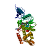
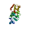
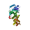
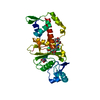
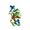
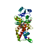
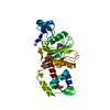
- PDB-1mjb: Crystal structure of yeast Esa1 histone acetyltransferase E338Q m... -
+ Open data

- Basic information
| Entry | Database: PDB / ID: 1mjb | ||||||
|---|---|---|---|---|---|---|---|
| Title | Crystal structure of yeast Esa1 histone acetyltransferase E338Q mutant complexed with acetyl coenzyme A | ||||||
 Components Components | Esa1 protein | ||||||
 Keywords Keywords |  TRANSFERASE / Esa1 / histone acetyltransferases / HAT / MYST TRANSFERASE / Esa1 / histone acetyltransferases / HAT / MYST | ||||||
| Function / homology |  Function and homology information Function and homology informationDNA Damage/Telomere Stress Induced Senescence / Sensing of DNA Double Strand Breaks / piccolo histone acetyltransferase complex / Recruitment and ATM-mediated phosphorylation of repair and signaling proteins at DNA double strand breaks / histone H4K16 acetyltransferase activity / peptide 2-hydroxyisobutyryltransferase activity / histone crotonyltransferase activity / DNA-templated transcription elongation / positive regulation of triglyceride biosynthetic process / histone H4 acetyltransferase activity ...DNA Damage/Telomere Stress Induced Senescence / Sensing of DNA Double Strand Breaks / piccolo histone acetyltransferase complex / Recruitment and ATM-mediated phosphorylation of repair and signaling proteins at DNA double strand breaks / histone H4K16 acetyltransferase activity / peptide 2-hydroxyisobutyryltransferase activity / histone crotonyltransferase activity / DNA-templated transcription elongation / positive regulation of triglyceride biosynthetic process / histone H4 acetyltransferase activity / rDNA heterochromatin formation / peptide N-acetyltransferase activity / NuA4 histone acetyltransferase complex / peptide-lysine-N-acetyltransferase activity / Estrogen-dependent gene expression / positive regulation of macroautophagy / histone acetyltransferase activity / histone acetyltransferase / Transferases; Acyltransferases; Transferring groups other than aminoacyl groups / positive regulation of transcription elongation by RNA polymerase II / transcription coregulator activity / nucleosome / regulation of cell cycle / DNA repair / negative regulation of DNA-templated transcription / DNA-templated transcription / chromatin / regulation of transcription by RNA polymerase II / positive regulation of transcription by RNA polymerase II / nucleusSimilarity search - Function | ||||||
| Biological species |  Saccharomyces cerevisiae (brewer's yeast) Saccharomyces cerevisiae (brewer's yeast) | ||||||
| Method | X-RAY DIFFRACTION / MOLECULAR REPLACEMENT / Resolution: 2.5 Å | ||||||
 Authors Authors | Yan, Y. / Harper, S. / Speicher, D. / Marmorstein, R. | ||||||
 Citation Citation | Journal: Nat.Struct.Biol. / Year: 2002 Title: The catalytic mechanism of the ESA1 histone acetyltransferase involves a self-acetylated intermediate. Authors: Yan, Y. / Harper, S. / Speicher, D.W. / Marmorstein, R. | ||||||
| History |
|
- Structure visualization
| Structure viewer | Molecule: MolmilJmol/JSmol |
|---|
- Downloads & links
-Download
| PDBx/mmCIF format | 1mjb.cif.gz | 69.7 KB | Display | PDBx/mmCIF format |
|---|---|---|---|---|
| PDB format | pdb1mjb.ent.gz | 56.5 KB | Display | PDB format |
| PDBx/mmJSON format | 1mjb.json.gz | Tree view | PDBx/mmJSON format | |
| Others |  Other downloads Other downloads |
-Validation report
| Arichive directory | https://data.pdbj.org/pub/pdb/validation_reports/mj/1mjbftp://data.pdbj.org/pub/pdb/validation_reports/mj/1mjb | HTTPS FTP |
|---|
-Related structure data
| Related structure data |  1mj9C  1mjaC  1fy7S S: Starting model for refinement C: citing same article ( |
|---|---|
| Similar structure data |


-Links
 PDBj
PDBj


- Assembly
| Deposited unit | 
| ||||||||
|---|---|---|---|---|---|---|---|---|---|
| 1 |
| ||||||||
| 2 | 
| ||||||||
| 3 | x 6
| ||||||||
| Unit cell |
|
-Components
| #1: Protein | Mass: 33387.430 Da / Num. of mol.: 1 Fragment: Histone acetyltransferase domain (Residues 160-445) Mutation: E338Q Source method: isolated from a genetically manipulated source Source: (gene. exp.) Saccharomyces cerevisiae (brewer's yeast)Gene: YOR244W / Plasmid: pRSET-A / Production host:  Escherichia coli (E. coli) / Strain (production host): BL21(lysS) / References: UniProt: Q08649 Escherichia coli (E. coli) / Strain (production host): BL21(lysS) / References: UniProt: Q08649 |
|---|---|
| #2: Chemical | ChemComp-ACO / Acetyl-CoA  Mass: 809.571 Da / Num. of mol.: 1 / Source method: obtained synthetically / Formula: C23H38N7O17P3S Mass: 809.571 Da / Num. of mol.: 1 / Source method: obtained synthetically / Formula: C23H38N7O17P3S |
| #3: Water | ChemComp-HOH / Water Mass: 18.015 Da / Num. of mol.: 90 / Source method: isolated from a natural source / Formula: H2O Mass: 18.015 Da / Num. of mol.: 90 / Source method: isolated from a natural source / Formula: H2O |
-Experimental details
-Experiment
| Experiment | Method: X-RAY DIFFRACTION / Number of used crystals: 1 |
|---|
- Sample preparation
| Crystal | Density Matthews: 3.75 Å3/Da / Density % sol: 67.17 % | ||||||||||||||||||||||||||||||
|---|---|---|---|---|---|---|---|---|---|---|---|---|---|---|---|---|---|---|---|---|---|---|---|---|---|---|---|---|---|---|---|
| Crystal grow | Temperature: 293 K / Method: vapor diffusion, hanging drop / pH: 6.5 Details: sodium cacodylate, ammonium sulfate, pH 6.5, VAPOR DIFFUSION, HANGING DROP, temperature 293K | ||||||||||||||||||||||||||||||
| Crystal grow | *PLUS Temperature: 20 ℃ | ||||||||||||||||||||||||||||||
| Components of the solutions | *PLUS
|
-Data collection
| Diffraction | Mean temperature: 93 K |
|---|---|
| Diffraction source | Source: ROTATING ANODE / Type: RIGAKU RU200 / Wavelength: 1.5418 Å |
| Detector | Type: RIGAKU RAXIS IV / Detector: IMAGE PLATE / Date: Aug 11, 2001 / Details: mirrors |
| Radiation | Monochromator: Yale mirrors / Protocol: SINGLE WAVELENGTH / Monochromatic (M) / Laue (L): M / Scattering type: x-ray |
| Radiation wavelength | Wavelength: 1.5418 Å / Relative weight: 1 |
| Reflection | Resolution: 2.5→20 Å / Num. all: 17458 / Num. obs: 17458 / % possible obs: 97 % / Observed criterion σ(F): 0 / Observed criterion σ(I): -3 / Redundancy: 9.3 % / Rmerge(I) obs: 0.056 / Rsym value: 0.056 / Net I/σ(I): 13.6 |
| Reflection shell | Resolution: 2.5→2.59 Å / Redundancy: 2.8 % / Rmerge(I) obs: 0.291 / Mean I/σ(I) obs: 3 / Num. unique all: 1701 / Rsym value: 0.291 / % possible all: 96.4 |
| Reflection | *PLUS Lowest resolution: 20 Å / Num. obs: 18056 / Num. measured all: 162442 / Rmerge(I) obs: 0.056 |
| Reflection shell | *PLUS Rmerge(I) obs: 0.291 |
- Processing
| Software |
| |||||||||||||||||||||||||
|---|---|---|---|---|---|---|---|---|---|---|---|---|---|---|---|---|---|---|---|---|---|---|---|---|---|---|
| Refinement | Method to determine structure: MOLECULAR REPLACEMENT Starting model: PDB entry 1FY7 Resolution: 2.5→20 Å / Isotropic thermal model: Isotropic / Cross valid method: THROUGHOUT / σ(F): 0 / σ(I): 0 / Stereochemistry target values: Engh & Huber
| |||||||||||||||||||||||||
| Refinement step | Cycle: LAST / Resolution: 2.5→20 Å
| |||||||||||||||||||||||||
| LS refinement shell | Resolution: 2.5→2.52 Å /
| |||||||||||||||||||||||||
| Xplor file |
| |||||||||||||||||||||||||
| Refinement | *PLUS Lowest resolution: 20 Å / % reflection Rfree: 10 % / Rfactor obs: 0.232 / Rfactor Rfree: 0.234 / Rfactor Rwork: 0.229 | |||||||||||||||||||||||||
| Solvent computation | *PLUS | |||||||||||||||||||||||||
| Displacement parameters | *PLUS | |||||||||||||||||||||||||
| Refine LS restraints | *PLUS
| |||||||||||||||||||||||||
| LS refinement shell | *PLUS Rfactor Rfree: 0.288 / Rfactor Rwork: 0.283 |
