 Movie
Movie Controller


+ Open data

- Basic information
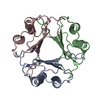
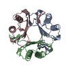
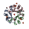
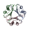
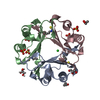
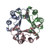
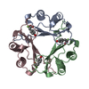
| Entry | Database: PDB / ID: 1mif | ||||||
|---|---|---|---|---|---|---|---|
| Title | MACROPHAGE MIGRATION INHIBITORY FACTOR (MIF) | ||||||
 Components Components | MACROPHAGE MIGRATION INHIBITORY FACTOR | ||||||
 Keywords Keywords | CYTOKINE / HORMONE / GLUTATHIONE BINDING PROTEIN | ||||||
| Function / homology |  Function and homology information Function and homology informationpositive regulation of prostaglandin secretion involved in immune response / positive regulation of myeloid leukocyte cytokine production involved in immune response / phenylpyruvate tautomerase / L-dopachrome isomerase / dopachrome isomerase activity / phenylpyruvate tautomerase activity / positive regulation of lipopolysaccharide-mediated signaling pathway / cytokine receptor binding / positive regulation of arachidonic acid secretion / negative regulation of myeloid cell apoptotic process ...positive regulation of prostaglandin secretion involved in immune response / positive regulation of myeloid leukocyte cytokine production involved in immune response / phenylpyruvate tautomerase / L-dopachrome isomerase / dopachrome isomerase activity / phenylpyruvate tautomerase activity / positive regulation of lipopolysaccharide-mediated signaling pathway / cytokine receptor binding / positive regulation of arachidonic acid secretion / negative regulation of myeloid cell apoptotic process / negative regulation of mature B cell apoptotic process / negative regulation of macrophage chemotaxis / positive regulation of chemokine (C-X-C motif) ligand 2 production / carboxylic acid metabolic process / prostaglandin biosynthetic process / negative regulation of protein metabolic process / regulation of macrophage activation / negative regulation of intrinsic apoptotic signaling pathway in response to DNA damage by p53 class mediator / chemoattractant activity / positive regulation of protein kinase A signaling / protein homotrimerization / negative regulation of cellular senescence / negative regulation of DNA damage response, signal transduction by p53 class mediator / DNA damage response, signal transduction by p53 class mediator / positive regulation of B cell proliferation / positive regulation of phosphorylation / Gene and protein expression by JAK-STAT signaling after Interleukin-12 stimulation / negative regulation of cell migration / positive regulation of cytokine production / cytokine activity / Cell surface interactions at the vascular wall / positive regulation of tumor necrosis factor production / cellular senescence / positive regulation of peptidyl-tyrosine phosphorylation / positive regulation of fibroblast proliferation / positive regulation of peptidyl-serine phosphorylation / secretory granule lumen / protease binding / vesicle / ficolin-1-rich granule lumen / cell surface receptor signaling pathway / positive regulation of ERK1 and ERK2 cascade / inflammatory response / negative regulation of gene expression / innate immune response / Neutrophil degranulation / positive regulation of cell population proliferation / negative regulation of apoptotic process / cell surface / extracellular space / extracellular exosome / extracellular region / nucleoplasm / identical protein binding / plasma membrane / cytosol / cytoplasmSimilarity search - Function | ||||||
| Biological species |  Homo sapiens (human) Homo sapiens (human) | ||||||
| Method | X-RAY DIFFRACTION / Resolution: 2.6 Å | ||||||
 Authors Authors | Sun, H.-W. / Lolis, E. | ||||||
 Citation Citation | Journal: Proc.Natl.Acad.Sci.USA / Year: 1996 Title: Crystal structure at 2.6-A resolution of human macrophage migration inhibitory factor. Authors: Sun, H.W. / Bernhagen, J. / Bucala, R. / Lolis, E. #1: Journal: To be PublishedTitle: The Subunit Structure of Human Migration Inhibitory Factor: Evidence for a Trimer Authors: Sun, H.-W. / Swope, M. / Cinquina, C. / Bedarkar, S. / Bernhagen, J. / Bucala, R. / Lolis, E. | ||||||
| History |
|
- Structure visualization
| Structure viewer | Molecule: MolmilJmol/JSmol |
|---|
- Downloads & links
-Download
| PDBx/mmCIF format | 1mif.cif.gz | 65.2 KB | Display | PDBx/mmCIF format |
|---|---|---|---|---|
| PDB format | pdb1mif.ent.gz | 49 KB | Display | PDB format |
| PDBx/mmJSON format | 1mif.json.gz | Tree view | PDBx/mmJSON format | |
| Others |  Other downloads Other downloads |
-Validation report
| Arichive directory | https://data.pdbj.org/pub/pdb/validation_reports/mi/1mifftp://data.pdbj.org/pub/pdb/validation_reports/mi/1mif | HTTPS FTP |
|---|
-Related structure data
| Similar structure data |
|---|


-Links
 PDBj
PDBj


- Assembly
| Deposited unit | 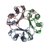
| ||||||||||||
|---|---|---|---|---|---|---|---|---|---|---|---|---|---|
| 1 |
| ||||||||||||
| Unit cell |
| ||||||||||||
| Noncrystallographic symmetry (NCS) | NCS oper:
|
-Components
| #1: Protein | / MIF Mass: 12459.227 Da / Num. of mol.: 3 Source method: isolated from a genetically manipulated source Source: (gene. exp.) Homo sapiens (human) / Plasmid: PET-11B / Species (production host): Escherichia coli / Production host:  Escherichia coli BL21(DE3) (bacteria) / Strain (production host): BL21(DE3) / References: UniProt: P14174 Escherichia coli BL21(DE3) (bacteria) / Strain (production host): BL21(DE3) / References: UniProt: P14174#2: Water | ChemComp-HOH / | Water Mass: 18.015 Da / Num. of mol.: 24 / Source method: isolated from a natural source / Formula: H2O Mass: 18.015 Da / Num. of mol.: 24 / Source method: isolated from a natural source / Formula: H2O |
|---|
-Experimental details
-Experiment
| Experiment | Method: X-RAY DIFFRACTION |
|---|
- Sample preparation
| Crystal | Density Matthews: 3.84 Å3/Da / Density % sol: 68 % | ||||||||||||||||||||||||||||||||||||||||
|---|---|---|---|---|---|---|---|---|---|---|---|---|---|---|---|---|---|---|---|---|---|---|---|---|---|---|---|---|---|---|---|---|---|---|---|---|---|---|---|---|---|
| Crystal grow | *PLUS pH: 7.5 / Method: vapor diffusion, hanging drop | ||||||||||||||||||||||||||||||||||||||||
| Components of the solutions | *PLUS
|
-Data collection
| Diffraction source | Source: ROTATING ANODE / Wavelength: 1.5418 |
|---|---|
| Detector | Type: SIEMENS-NICOLET X100 / Detector: AREA DETECTOR / Date: Jun 14, 1995 |
| Radiation | Monochromatic (M) / Laue (L): M / Scattering type: x-ray |
| Radiation wavelength | Wavelength: 1.5418 Å / Relative weight: 1 |
| Reflection | Num. obs: 18101 / % possible obs: 82.1 % / Observed criterion σ(I): 2 / Redundancy: 2.6 % / Rmerge(I) obs: 0.053 |
- Processing
| Software |
| ||||||||||||||||||||||||||||||||||||||||||||||||||||||||||||
|---|---|---|---|---|---|---|---|---|---|---|---|---|---|---|---|---|---|---|---|---|---|---|---|---|---|---|---|---|---|---|---|---|---|---|---|---|---|---|---|---|---|---|---|---|---|---|---|---|---|---|---|---|---|---|---|---|---|---|---|---|---|
| Refinement | Resolution: 2.6→5 Å / σ(F): 2 /
| ||||||||||||||||||||||||||||||||||||||||||||||||||||||||||||
| Refinement step | Cycle: LAST / Resolution: 2.6→5 Å
| ||||||||||||||||||||||||||||||||||||||||||||||||||||||||||||
| Refine LS restraints |
| ||||||||||||||||||||||||||||||||||||||||||||||||||||||||||||
| Software | *PLUS Name: X-PLOR / Classification: refinement | ||||||||||||||||||||||||||||||||||||||||||||||||||||||||||||
| Refinement | *PLUS | ||||||||||||||||||||||||||||||||||||||||||||||||||||||||||||
| Solvent computation | *PLUS | ||||||||||||||||||||||||||||||||||||||||||||||||||||||||||||
| Displacement parameters | *PLUS | ||||||||||||||||||||||||||||||||||||||||||||||||||||||||||||
| Refine LS restraints | *PLUS Type: x_angle_deg / Dev ideal: 1.8 |
