 Movie
Movie Controller


+ Open data

- Basic information
| Entry | Database: PDB / ID: 1mhd | ||||||
|---|---|---|---|---|---|---|---|
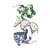
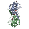
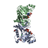
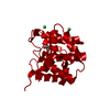
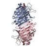
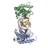
| Title | CRYSTAL STRUCTURE OF A SMAD MH1 DOMAIN BOUND TO DNA | ||||||
 Components Components |
| ||||||
 Keywords Keywords | COMPLEX (TRANSCRIPTION ACTIVATOR/DNA) / COMPLEX (TRANSCRIPTION ACTIVATOR-DNA) / SMAD3 MH1 / SMAD BINDING ELEMENT /  DNA / COMPLEX (TRANSCRIPTION ACTIVATOR-DNA) complex DNA / COMPLEX (TRANSCRIPTION ACTIVATOR-DNA) complex | ||||||
| Function / homology |  Function and homology information Function and homology informationnuclear mineralocorticoid receptor binding / negative regulation of lung blood pressure / regulation of miRNA transcription / positive regulation of transforming growth factor beta3 production / transdifferentiation / sterol response element binding / paraxial mesoderm morphogenesis / SMAD4 MH2 Domain Mutants in Cancer / SMAD2/3 MH2 Domain Mutants in Cancer / nodal signaling pathway ...nuclear mineralocorticoid receptor binding / negative regulation of lung blood pressure / regulation of miRNA transcription / positive regulation of transforming growth factor beta3 production / transdifferentiation / sterol response element binding / paraxial mesoderm morphogenesis / SMAD4 MH2 Domain Mutants in Cancer / SMAD2/3 MH2 Domain Mutants in Cancer / nodal signaling pathway / regulation of striated muscle tissue development / SMAD protein complex / immune system development / co-SMAD binding / heteromeric SMAD protein complex / regulation of transforming growth factor beta2 production / RUNX3 regulates BCL2L11 (BIM) transcription / DEAD/H-box RNA helicase binding / bHLH transcription factor binding / pericardium development / FOXO-mediated transcription of cell cycle genes / SMAD2/3 Phosphorylation Motif Mutants in Cancer / TGFBR1 KD Mutants in Cancer / positive regulation of chondrocyte differentiation / negative regulation of osteoblast proliferation / regulation of transforming growth factor beta receptor signaling pathway / embryonic foregut morphogenesis / negative regulation of wound healing / positive regulation of extracellular matrix assembly / lens fiber cell differentiation / transforming growth factor beta receptor binding / primary miRNA processing / Germ layer formation at gastrulation / nuclear glucocorticoid receptor binding / Formation of definitive endoderm / endoderm development / embryonic pattern specification / activin receptor signaling pathway / signal transduction involved in regulation of gene expression / Signaling by Activin / SMAD protein signal transduction / Formation of axial mesoderm / cell-cell junction organization / Signaling by NODAL / regulation of epithelial cell proliferation / embryonic cranial skeleton morphogenesis / I-SMAD binding / Interleukin-37 signaling / response to angiotensin / positive regulation of positive chemotaxis / osteoblast development / NOTCH4 Intracellular Domain Regulates Transcription / RUNX3 regulates CDKN1A transcription / negative regulation of cardiac muscle hypertrophy in response to stress / nuclear inner membrane / ureteric bud development / DNA-binding transcription repressor activity / adrenal gland development / negative regulation of fat cell differentiation / negative regulation of cytosolic calcium ion concentration / heart looping / TGF-beta receptor signaling activates SMADs / positive regulation of focal adhesion assembly / R-SMAD binding / thyroid gland development / mesoderm formation / developmental growth / regulation of immune response / anatomical structure morphogenesis / phosphatase binding / negative regulation of osteoblast differentiation / cis-regulatory region sequence-specific DNA binding / FOXO-mediated transcription of oxidative stress, metabolic and neuronal genes / positive regulation of bone mineralization / somitogenesis / positive regulation of epithelial to mesenchymal transition / SARS-CoV-1 targets host intracellular signalling and regulatory pathways / extrinsic apoptotic signaling pathway / JNK cascade / positive regulation of stress fiber assembly / cellular response to transforming growth factor beta stimulus / collagen binding / T cell activation / Downregulation of TGF-beta receptor signaling / transforming growth factor beta receptor signaling pathway / transcription corepressor binding / negative regulation of miRNA transcription / liver development / ubiquitin binding / positive regulation of interleukin-1 beta production / promoter-specific chromatin binding / nuclear receptor binding / cellular response to glucose stimulus / Downregulation of SMAD2/3:SMAD4 transcriptional activity / SMAD2/SMAD3:SMAD4 heterotrimer regulates transcription / wound healing / negative regulation of protein catabolic process / negative regulation of cell growth / chromatin DNA binding / cellular response to virusSimilarity search - Function | ||||||
| Biological species |  Homo sapiens (human) Homo sapiens (human) | ||||||
| Method | X-RAY DIFFRACTION / MIR / Resolution: 2.8 Å | ||||||
 Authors Authors | Shi, Y. | ||||||
 Citation Citation | Journal: Cell(Cambridge,Mass.) / Year: 1998 Title: Crystal structure of a Smad MH1 domain bound to DNA: insights on DNA binding in TGF-beta signaling. Authors: Shi, Y. / Wang, Y.F. / Jayaraman, L. / Yang, H. / Massague, J. / Pavletich, N.P. | ||||||
| History |
|
- Structure visualization
| Structure viewer | Molecule: MolmilJmol/JSmol |
|---|
- Downloads & links
-Download
| PDBx/mmCIF format | 1mhd.cif.gz | 74.7 KB | Display | PDBx/mmCIF format |
|---|---|---|---|---|
| PDB format | pdb1mhd.ent.gz | 58.5 KB | Display | PDB format |
| PDBx/mmJSON format | 1mhd.json.gz | Tree view | PDBx/mmJSON format | |
| Others |  Other downloads Other downloads |
-Validation report
| Arichive directory | https://data.pdbj.org/pub/pdb/validation_reports/mh/1mhdftp://data.pdbj.org/pub/pdb/validation_reports/mh/1mhd | HTTPS FTP |
|---|
-Related structure data
| Similar structure data |
|---|


-Links
 PDBj
PDBj


- Assembly
| Deposited unit | 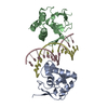
| ||||||||
|---|---|---|---|---|---|---|---|---|---|
| 1 |
| ||||||||
| Unit cell |
|
-Components
| #1: DNA chain | Mass: 3959.612 Da / Num. of mol.: 1 / Source method: obtained synthetically | ||
|---|---|---|---|
| #2: DNA chain | Mass: 4294.814 Da / Num. of mol.: 1 / Source method: obtained synthetically | ||
| #3: Protein | Mothers against decapentaplegic homolog 3 / SMAD N-DOMAIN Mass: 15539.169 Da / Num. of mol.: 2 / Fragment: MH1 DOMAIN, RESIDUES 1 - 144 Source method: isolated from a genetically manipulated source Source: (gene. exp.) Homo sapiens (human) / Cellular location: CYTOPLASM / Gene: SMAD / Plasmid: PGEX / Species (production host): Escherichia coli / Cellular location (production host): CYTOPLASM / Production host:  Escherichia coli BL21(DE3) (bacteria) / Strain (production host): BL21 (DE3) / References: UniProt: P84022 Escherichia coli BL21(DE3) (bacteria) / Strain (production host): BL21 (DE3) / References: UniProt: P84022#4: Water | ChemComp-HOH / | Water Mass: 18.015 Da / Num. of mol.: 24 / Source method: isolated from a natural source / Formula: H2O Mass: 18.015 Da / Num. of mol.: 24 / Source method: isolated from a natural source / Formula: H2O |
-Experimental details
-Experiment
| Experiment | Method: X-RAY DIFFRACTION / Number of used crystals: 2 |
|---|
- Sample preparation
| Crystal | Density Matthews: 2.43 Å3/Da / Density % sol: 36 % | ||||||||||||||||||||||||||||||||||||||||||
|---|---|---|---|---|---|---|---|---|---|---|---|---|---|---|---|---|---|---|---|---|---|---|---|---|---|---|---|---|---|---|---|---|---|---|---|---|---|---|---|---|---|---|---|
| Crystal grow | pH: 5.6 / Details: pH 5.6 | ||||||||||||||||||||||||||||||||||||||||||
| Crystal | *PLUS | ||||||||||||||||||||||||||||||||||||||||||
| Crystal grow | *PLUS pH: 5.5 / Method: vapor diffusion, hanging drop | ||||||||||||||||||||||||||||||||||||||||||
| Components of the solutions | *PLUS
|
-Data collection
| Diffraction | Mean temperature: 300 K |
|---|---|
| Diffraction source | Source: ROTATING ANODE / Type: RIGAKU / Wavelength: 1.5418 |
| Detector | Type: RIGAKU RAXIS IIC / Detector: IMAGE PLATE / Date: May 5, 1998 / Details: MIRRORS |
| Radiation | Monochromator: NI FILTER / Monochromatic (M) / Laue (L): M / Scattering type: x-ray |
| Radiation wavelength | Wavelength: 1.5418 Å / Relative weight: 1 |
| Reflection | Resolution: 2.8→20 Å / Num. obs: 9228 / % possible obs: 97 % / Observed criterion σ(I): 2 / Redundancy: 4.3 % / Rsym value: 0.053 / Net I/σ(I): 33 |
| Reflection shell | Resolution: 2.8→2.9 Å / Mean I/σ(I) obs: 9 / Rsym value: 0.162 / % possible all: 96.5 |
| Reflection | *PLUS Num. measured all: 39338 / Rmerge(I) obs: 0.053 |
| Reflection shell | *PLUS % possible obs: 96.5 % / Rmerge(I) obs: 0.162 |
- Processing
| Software |
| ||||||||||||||||||||||||||||||||||||||||||||||||||||||||||||
|---|---|---|---|---|---|---|---|---|---|---|---|---|---|---|---|---|---|---|---|---|---|---|---|---|---|---|---|---|---|---|---|---|---|---|---|---|---|---|---|---|---|---|---|---|---|---|---|---|---|---|---|---|---|---|---|---|---|---|---|---|---|
| Refinement | Method to determine structure: MIR / Resolution: 2.8→8 Å / Cross valid method: R FACTOR / σ(F): 2
| ||||||||||||||||||||||||||||||||||||||||||||||||||||||||||||
| Refinement step | Cycle: LAST / Resolution: 2.8→8 Å
| ||||||||||||||||||||||||||||||||||||||||||||||||||||||||||||
| Refine LS restraints |
| ||||||||||||||||||||||||||||||||||||||||||||||||||||||||||||
| LS refinement shell | Resolution: 2.8→2.92 Å / Total num. of bins used: 8
| ||||||||||||||||||||||||||||||||||||||||||||||||||||||||||||
| Software | *PLUS Name: X-PLOR / Version: 3.1 / Classification: refinement | ||||||||||||||||||||||||||||||||||||||||||||||||||||||||||||
| LS refinement shell | *PLUS Rfactor Rwork: 0.34 |
