 Movie
Movie Controller

[English] 日本語
 Yorodumi
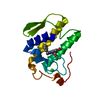
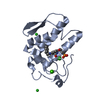
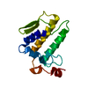
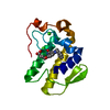
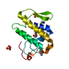
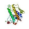
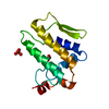
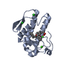
- PDB-1mh7: Crystal Structure of a Calcium-Free Isoform of Phospholipase A2 f... -
+ Open data

- Basic information
| Entry | Database: PDB / ID: 1mh7 | ||||||
|---|---|---|---|---|---|---|---|
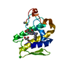
| Title | Crystal Structure of a Calcium-Free Isoform of Phospholipase A2 from Naja naja sagittifera at 2.0 A Resolution | ||||||
 Components Components | PHOSPHOLIPASE A2 | ||||||
 Keywords Keywords | HYDROLASE / PHOSPHOLIPASE / ENZYME / PHOSPHOLIPIDS | ||||||
| Function / homology |  Function and homology informationphospholipase A2 activity / phospholipase A2 / arachidonic acid secretion / phospholipid metabolic process / lipid catabolic process / calcium ion binding / extracellular region Function and homology informationphospholipase A2 activity / phospholipase A2 / arachidonic acid secretion / phospholipid metabolic process / lipid catabolic process / calcium ion binding / extracellular regionSimilarity search - Function | ||||||
| Biological species |  Naja sagittifera (cobra) Naja sagittifera (cobra) | ||||||
| Method | X-RAY DIFFRACTION / MOLECULAR REPLACEMENT / Resolution: 2 Å | ||||||
 Authors Authors | Jabeen, T. / Jasti, J. / Singh, R.K. / Sujata, S. / Singh, T.P. | ||||||
 Citation Citation | Journal: To be Published Title: Crystal Structure of a Calcium-Free Isoform of Phospholipase A2 from Naja naja sagittifera at 2.0 A Resolution Authors: Jabeen, T. / Jasti, J. / Singh, R.K. / Sujata, S. / Singh, T.P. | ||||||
| History |
|
- Structure visualization
| Structure viewer | Molecule: MolmilJmol/JSmol |
|---|
- Downloads & links
-Download
| PDBx/mmCIF format | 1mh7.cif.gz | 32.5 KB | Display | PDBx/mmCIF format |
|---|---|---|---|---|
| PDB format | pdb1mh7.ent.gz | 24.8 KB | Display | PDB format |
| PDBx/mmJSON format | 1mh7.json.gz | Tree view | PDBx/mmJSON format | |
| Others |  Other downloads Other downloads |
-Validation report
| Arichive directory | https://data.pdbj.org/pub/pdb/validation_reports/mh/1mh7ftp://data.pdbj.org/pub/pdb/validation_reports/mh/1mh7 | HTTPS FTP |
|---|
-Related structure data
| Related structure data |  1lfj S: Starting model for refinement |
|---|---|
| Similar structure data |


-Links
 PDBj
PDBj

- Assembly
| Deposited unit | 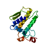
| ||||||||
|---|---|---|---|---|---|---|---|---|---|
| 1 |
| ||||||||
| Unit cell |
|
-Components
| #1: Protein | Mass: 13319.810 Da / Num. of mol.: 1 / Source method: isolated from a natural source / Source: (natural) Naja sagittifera (cobra) / Secretion: VENOM / References: UniProt: P60043, phospholipase A2 |
|---|---|
| #2: Water | ChemComp-HOH / Water Mass: 18.015 Da / Num. of mol.: 74 / Source method: isolated from a natural source / Formula: H2O Mass: 18.015 Da / Num. of mol.: 74 / Source method: isolated from a natural source / Formula: H2O |
-Experimental details
-Experiment
| Experiment | Method: X-RAY DIFFRACTION / Number of used crystals: 1 |
|---|
- Sample preparation
| Crystal | Density Matthews: 2.26 Å3/Da / Density % sol: 45.49 % |
|---|---|
| Crystal grow | Temperature: 293 K / Method: vapor diffusion, sitting drop / pH: 6.5 Details: 10 mM SODIUM PHOSPHATE, 2mM CaCl2, 28% ETHANOL, pH 6.5, VAPOR DIFFUSION, SITTING DROP, temperature 293K |
-Data collection
| Diffraction | Mean temperature: 298 K |
|---|---|
| Diffraction source | Source: ROTATING ANODE / Type: RIGAKU RU200 / Wavelength: 1.5418 Å |
| Detector | Type: MARRESEARCH / Detector: IMAGE PLATE / Date: Jun 1, 2002 / Details: MONOCHRMOATOR |
| Radiation | Monochromator: GRAPHITE / Protocol: SINGLE WAVELENGTH / Monochromatic (M) / Laue (L): M / Scattering type: x-ray |
| Radiation wavelength | Wavelength: 1.5418 Å / Relative weight: 1 |
| Reflection | Resolution: 2→20 Å / Num. obs: 7717 / % possible obs: 97 % / Observed criterion σ(F): 0 / Observed criterion σ(I): -3 / Redundancy: 6.4 % / Biso Wilson estimate: 21.8 Å2 / Rmerge(I) obs: 0.084 / Rsym value: 0.083 / Net I/σ(I): 22.1 |
| Reflection shell | Resolution: 2→2.07 Å / Redundancy: 4.6 % / Rmerge(I) obs: 0.23 / Mean I/σ(I) obs: 2.7 / Num. unique all: 494 / Rsym value: 0.237 / % possible all: 82 |
- Processing
| Software |
| ||||||||||||||||||||||||||||||||||||||||||||||||||||||||||||||||||||||||||||||||
|---|---|---|---|---|---|---|---|---|---|---|---|---|---|---|---|---|---|---|---|---|---|---|---|---|---|---|---|---|---|---|---|---|---|---|---|---|---|---|---|---|---|---|---|---|---|---|---|---|---|---|---|---|---|---|---|---|---|---|---|---|---|---|---|---|---|---|---|---|---|---|---|---|---|---|---|---|---|---|---|---|---|
| Refinement | Method to determine structure: MOLECULAR REPLACEMENT Starting model: 1LFJ 1lfj Resolution: 2→20 Å / Rfactor Rfree error: 0.011 / Isotropic thermal model: RESTRAINED / Cross valid method: THROUGHOUT / σ(F): 0 / σ(I): 0 / Stereochemistry target values: Engh & Huber
| ||||||||||||||||||||||||||||||||||||||||||||||||||||||||||||||||||||||||||||||||
| Solvent computation | Solvent model: FLAT MODEL / Bsol: 34.06 Å2 / ksol: 0.265159 e/Å3 | ||||||||||||||||||||||||||||||||||||||||||||||||||||||||||||||||||||||||||||||||
| Displacement parameters | Biso mean: 25.2 Å2 | ||||||||||||||||||||||||||||||||||||||||||||||||||||||||||||||||||||||||||||||||
| Refine analyze |
| ||||||||||||||||||||||||||||||||||||||||||||||||||||||||||||||||||||||||||||||||
| Refinement step | Cycle: LAST / Resolution: 2→20 Å
| ||||||||||||||||||||||||||||||||||||||||||||||||||||||||||||||||||||||||||||||||
| Refine LS restraints |
| ||||||||||||||||||||||||||||||||||||||||||||||||||||||||||||||||||||||||||||||||
| LS refinement shell | Resolution: 2→2.13 Å / Rfactor Rfree error: 0.033 / Total num. of bins used: 6
| ||||||||||||||||||||||||||||||||||||||||||||||||||||||||||||||||||||||||||||||||
| Xplor file |
|
