 Movie
Movie Controller

[English] 日本語
 Yorodumi
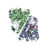
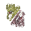
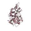
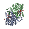
- PDB-1mg5: Crystal structure of Drosophila melanogaster alcohol dehydrogenas... -
+ Open data

- Basic information
| Entry | Database: PDB / ID: 1mg5 | ||||||
|---|---|---|---|---|---|---|---|
| Title | Crystal structure of Drosophila melanogaster alcohol dehydrogenase complexed with NADH and acetate at 1.6 A | ||||||
 Components Components | alcohol dehydrogenase | ||||||
 Keywords Keywords | OXIDOREDUCTASE / SDR / ADH / Drosophila melanogaster / NADH / acetate | ||||||
| Function / homology |  Function and homology information Function and homology informationalcohol catabolic process / ethanol metabolic process / acetaldehyde dehydrogenase (acetylating) activity / alcohol metabolic process / alcohol dehydrogenase (NAD+) activity / acetaldehyde metabolic process / : / behavioral response to ethanol / : / alcohol dehydrogenase ...alcohol catabolic process / ethanol metabolic process / acetaldehyde dehydrogenase (acetylating) activity / alcohol metabolic process / alcohol dehydrogenase (NAD+) activity / acetaldehyde metabolic process / : / behavioral response to ethanol / : / alcohol dehydrogenase / oxidoreductase activity / protein homodimerization activity / protein-containing complex / cytosolSimilarity search - Function | ||||||
| Biological species |  Drosophila melanogaster (fruit fly) Drosophila melanogaster (fruit fly) | ||||||
| Method | X-RAY DIFFRACTION / MOLECULAR REPLACEMENT / Resolution: 1.63 Å | ||||||
 Authors Authors | Benach, J. / Atrian, S. / Gonzalez-Duarte, R. / Ladenstein, R. | ||||||
 Citation Citation | Journal: J.Mol.Biol. / Year: 2005 Title: Drosophila alcohol dehydrogenase: acetate-enzyme interactions and novel insights into the effects of electrostatics on catalysis Authors: Benach, J. / Winberg, J.O. / Svendsen, J.S. / Atrian, S. / Gonzalez-Duarte, R. / Ladenstein, R. #1: Journal: J.Mol.Biol. / Year: 1998Title: The refined crystal structure of Drosophila lebanonensis alcohol dehydrogenase at 1.9 A resolution Authors: Benach, J. / Atrian, S. / Gonzalez-Duarte, R. / Ladenstein, R. #2: Journal: J.Mol.Biol. / Year: 1999Title: The catalytic reaction and inhibition mechanism of Drosophila alcohol dehydrogenase: Observation of an enzyme-bound NAD-ketone adduct at 1.4 A resolution by X-ray crystallography Authors: Benach, J. / Atrian, S. / Gonzalez-Duarte, R. / Ladenstein, R. | ||||||
| History |
| ||||||
| Remark 999 | sequence Since the protein was purified directly from Drosophila melanogaster ADH. The first ...sequence Since the protein was purified directly from Drosophila melanogaster ADH. The first residue: Met1 is cleaved off in the matured Drosophila melanogaster ADH-S enzyme. For this reason it is not present in the crystal structure. |
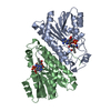
- Structure visualization
| Structure viewer | Molecule: MolmilJmol/JSmol |
|---|
- Downloads & links
-Download
| PDBx/mmCIF format | 1mg5.cif.gz | 129.8 KB | Display | PDBx/mmCIF format |
|---|---|---|---|---|
| PDB format | pdb1mg5.ent.gz | 99.2 KB | Display | PDB format |
| PDBx/mmJSON format | 1mg5.json.gz | Tree view | PDBx/mmJSON format | |
| Others |  Other downloads Other downloads |
-Validation report
| Arichive directory | https://data.pdbj.org/pub/pdb/validation_reports/mg/1mg5ftp://data.pdbj.org/pub/pdb/validation_reports/mg/1mg5 | HTTPS FTP |
|---|
-Related structure data
| Related structure data |  1a4uS S: Starting model for refinement |
|---|---|
| Similar structure data |


-Links
 PDBj
PDBj


- Assembly
| Deposited unit | 
| ||||||||
|---|---|---|---|---|---|---|---|---|---|
| 1 |
| ||||||||
| Unit cell |
|
-Components
| #1: Protein | Mass: 27656.834 Da / Num. of mol.: 2 / Source method: isolated from a natural source / Details: ADH-Slow allele / Source: (natural) Drosophila melanogaster (fruit fly) / References: UniProt: P00334, alcohol dehydrogenase#2: Chemical | Acetate  Mass: 59.044 Da / Num. of mol.: 2 / Source method: obtained synthetically / Formula: C2H3O2 Mass: 59.044 Da / Num. of mol.: 2 / Source method: obtained synthetically / Formula: C2H3O2#3: Chemical | Nicotinamide adenine dinucleotide  Mass: 665.441 Da / Num. of mol.: 2 / Source method: obtained synthetically / Formula: C21H29N7O14P2 Mass: 665.441 Da / Num. of mol.: 2 / Source method: obtained synthetically / Formula: C21H29N7O14P2#4: Water | ChemComp-HOH / | Water Mass: 18.015 Da / Num. of mol.: 822 / Source method: isolated from a natural source / Formula: H2O Mass: 18.015 Da / Num. of mol.: 822 / Source method: isolated from a natural source / Formula: H2O |
|---|
-Experimental details
-Experiment
| Experiment | Method: X-RAY DIFFRACTION / Number of used crystals: 1 |
|---|
- Sample preparation
| Crystal | Density Matthews: 2.26 Å3/Da / Density % sol: 45.68 % | ||||||||||||||||||||||||||||||
|---|---|---|---|---|---|---|---|---|---|---|---|---|---|---|---|---|---|---|---|---|---|---|---|---|---|---|---|---|---|---|---|
| Crystal grow | Temperature: 277 K / Method: vapor diffusion, sitting drop / pH: 8.5 Details: 0.1 M TRIS/HCl, 30% PEG4000, 0.2M NaAcetate trihydrate, pH 8.5, VAPOR DIFFUSION, SITTING DROP, temperature 277K | ||||||||||||||||||||||||||||||
| Crystal grow | *PLUS Temperature: 277 K / pH: 8.6 / Method: vapor diffusion | ||||||||||||||||||||||||||||||
| Components of the solutions | *PLUS
|
-Data collection
| Diffraction | Mean temperature: 100 K |
|---|---|
| Diffraction source | Source: ROTATING ANODE / Type: SIEMENS / Wavelength: 1.5418 Å |
| Detector | Type: MARRESEARCH / Detector: IMAGE PLATE / Date: Feb 10, 2000 |
| Radiation | Protocol: SINGLE WAVELENGTH / Monochromatic (M) / Laue (L): M / Scattering type: x-ray |
| Radiation wavelength | Wavelength: 1.5418 Å / Relative weight: 1 |
| Reflection | Resolution: 1.63→40 Å / Num. obs: 60633 / % possible obs: 96.7 % / Observed criterion σ(F): 0 / Observed criterion σ(I): -3 / Redundancy: 7.5 % / Biso Wilson estimate: 15.3 Å2 / Rmerge(I) obs: 0.082 / Net I/σ(I): 19.7 |
| Reflection shell | Resolution: 1.63→1.68 Å / Rmerge(I) obs: 0.36 / % possible all: 73.3 |
| Reflection | *PLUS Num. measured all: 438968 |
| Reflection shell | *PLUS % possible obs: 73.3 % / Rmerge(I) obs: 0.36 |
- Processing
| Software |
| |||||||||||||||||||||
|---|---|---|---|---|---|---|---|---|---|---|---|---|---|---|---|---|---|---|---|---|---|---|
| Refinement | Method to determine structure: MOLECULAR REPLACEMENT Starting model: 1a4u Resolution: 1.63→20 Å / Isotropic thermal model: isotropic / Cross valid method: free R-factor / σ(F): 0 / σ(I): 0 / Stereochemistry target values: Engh & Huber
| |||||||||||||||||||||
| Displacement parameters | Biso mean: 14.6 Å2 | |||||||||||||||||||||
| Refine analyze | Luzzati coordinate error obs: 0.16 Å / Luzzati d res low obs: 5 Å | |||||||||||||||||||||
| Refinement step | Cycle: LAST / Resolution: 1.63→20 Å
| |||||||||||||||||||||
| Refine LS restraints |
| |||||||||||||||||||||
| Refinement | *PLUS | |||||||||||||||||||||
| Solvent computation | *PLUS | |||||||||||||||||||||
| Displacement parameters | *PLUS | |||||||||||||||||||||
| Refine LS restraints | *PLUS
|
