 Movie
Movie Controller

[English] 日本語
 Yorodumi
- PDB-1m9i: Crystal Structure Of Phosphorylation-Mimicking Mutant T356D Of An... -
+ Open data

- Basic information
| Entry | Database: PDB / ID: 1m9i | ||||||
|---|---|---|---|---|---|---|---|
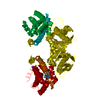
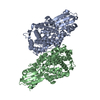
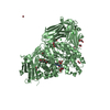
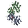
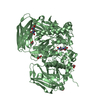
| Title | Crystal Structure Of Phosphorylation-Mimicking Mutant T356D Of Annexin VI | ||||||
 Components Components | Annexin VI | ||||||
 Keywords Keywords | LIPID BINDING PROTEIN / ANNEXIN / CALCIUM-BINDING / MEMBRANE-BINDING / PHOSPHORYLATION / MUTANT T356D | ||||||
| Function / homology |  Function and homology information Function and homology informationnegative regulation of sequestering of calcium ion / growth plate cartilage chondrocyte differentiation / chondroitin sulfate binding / mitochondrial calcium ion homeostasis / regulation of muscle contraction / plasma membrane repair / calcium-dependent phospholipid binding / neural crest cell migration / cholesterol binding / phosphatidylserine binding ...negative regulation of sequestering of calcium ion / growth plate cartilage chondrocyte differentiation / chondroitin sulfate binding / mitochondrial calcium ion homeostasis / regulation of muscle contraction / plasma membrane repair / calcium-dependent phospholipid binding / neural crest cell migration / cholesterol binding / phosphatidylserine binding / ligand-gated monoatomic ion channel activity / Smooth Muscle Contraction / monoatomic ion transmembrane transport / apoptotic signaling pathway / calcium channel activity / calcium-dependent protein binding / actin filament binding / melanosome / late endosome membrane / collagen-containing extracellular matrix / lysosomal membrane / focal adhesion / lipid binding / calcium ion binding / GTP binding / perinuclear region of cytoplasm / mitochondrion / extracellular exosome / membrane / identical protein binding / cytoplasmSimilarity search - Function | ||||||
| Biological species |  Homo sapiens (human) Homo sapiens (human) | ||||||
| Method | X-RAY DIFFRACTION / SYNCHROTRON / MOLECULAR REPLACEMENT / Resolution: 2.65 Å | ||||||
 Authors Authors | Freye-Minks, C. / Kretsinger, R.H. / Creutz, C.E. | ||||||
 Citation Citation | Journal: Biochemistry / Year: 2003 Title: Structural and Dynamic Changes in Human Annexin VI Induced by a Phosphorylation-Mimicking Mutation, T356D Authors: Freye-Minks, C. / Kretsinger, R.H. / Creutz, C.E. #1: Journal: J.Mol.Biol. / Year: 1996Title: The structure of recombinant human annexin VI in crystals and membrane-bound Authors: Benz, J. / Bergner, A. / Hofmann, A. / Demange, P. / Gottig, P. / Liemann, S. / Huber, R. / Voges, D. #2: Journal: BIOCHIM.BIOPHYS.ACTA / Year: 1998Title: Crystal structure of bovine annexin VI in a calcium-bound state Authors: Avila-Sakar, A.J. / Creutz, C.E. / Kretsinger, R.H. | ||||||
| History |
|
- Structure visualization
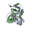
| Structure viewer | Molecule: MolmilJmol/JSmol |
|---|
- Downloads & links
-Download
| PDBx/mmCIF format | 1m9i.cif.gz | 145.9 KB | Display | PDBx/mmCIF format |
|---|---|---|---|---|
| PDB format | pdb1m9i.ent.gz | 114 KB | Display | PDB format |
| PDBx/mmJSON format | 1m9i.json.gz | Tree view | PDBx/mmJSON format | |
| Others |  Other downloads Other downloads |
-Validation report
| Arichive directory | https://data.pdbj.org/pub/pdb/validation_reports/m9/1m9iftp://data.pdbj.org/pub/pdb/validation_reports/m9/1m9i | HTTPS FTP |
|---|
-Related structure data
| Related structure data | |
|---|---|
| Similar structure data |


-Links
 PDBj
PDBj


- Assembly
| Deposited unit | 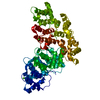
| ||||||||
|---|---|---|---|---|---|---|---|---|---|
| 1 |
| ||||||||
| Unit cell |
|
-Components
| #1: Protein | / Lipocortin VI / P68 / P70 / PROTEIN III / Chromobindin 20 / 67 kDa calelectrin / Calphobindin-II / CPB-II Mass: 75849.055 Da / Num. of mol.: 1 / Mutation: T356D Source method: isolated from a genetically manipulated source Source: (gene. exp.) Homo sapiens (human) / Gene: ANX6 / Plasmid: YepDB60 / Production host:  Saccharomyces cerevisiae (brewer's yeast) / Strain (production host): DB334 / References: UniProt: P08133 Saccharomyces cerevisiae (brewer's yeast) / Strain (production host): DB334 / References: UniProt: P08133 | ||
|---|---|---|---|
| #2: Chemical | ChemComp-CA /   Mass: 40.078 Da / Num. of mol.: 5 / Source method: obtained synthetically / Formula: Ca Mass: 40.078 Da / Num. of mol.: 5 / Source method: obtained synthetically / Formula: Ca#3: Water | ChemComp-HOH / | Water Mass: 18.015 Da / Num. of mol.: 148 / Source method: isolated from a natural source / Formula: H2O Mass: 18.015 Da / Num. of mol.: 148 / Source method: isolated from a natural source / Formula: H2O |
-Experimental details
-Experiment
| Experiment | Method: X-RAY DIFFRACTION / Number of used crystals: 1 |
|---|
- Sample preparation
| Crystal | Density Matthews: 3.11 Å3/Da / Density % sol: 60.51 % | ||||||||||||||||||||||||
|---|---|---|---|---|---|---|---|---|---|---|---|---|---|---|---|---|---|---|---|---|---|---|---|---|---|
| Crystal grow | Temperature: 298 K / Method: vapor diffusion / pH: 7.4 Details: 0.5M ammonium sulfate, 0.1M imidazole, pH 7.4, Vapor diffusion, temperature 298.0K | ||||||||||||||||||||||||
| Crystal grow | *PLUS Method: vapor diffusion, hanging drop | ||||||||||||||||||||||||
| Components of the solutions | *PLUS
|
-Data collection
| Diffraction | Mean temperature: 100 K |
|---|---|
| Diffraction source | Source: SYNCHROTRON / Site: NSLS  / Beamline: X4A / Wavelength: 0.971 Å / Beamline: X4A / Wavelength: 0.971 Å |
| Detector | Type: ADSC QUANTUM 4 / Detector: CCD / Date: Aug 16, 2001 / Details: Mirrors |
| Radiation | Monochromator: SAGITALLY FOCUSED Si(111) / Protocol: SINGLE WAVELENGTH / Monochromatic (M) / Laue (L): M / Scattering type: x-ray |
| Radiation wavelength | Wavelength: 0.971 Å / Relative weight: 1 |
| Reflection | Resolution: 2.5→50 Å / Num. obs: 32036 / % possible obs: 97.2 % / Observed criterion σ(F): 0 / Observed criterion σ(I): 0 / Redundancy: 5.6 % / Biso Wilson estimate: 34.7 Å2 / Rmerge(I) obs: 0.089 / Rsym value: 0.089 / Net I/σ(I): 22.8 |
| Reflection shell | Resolution: 2.5→2.59 Å / Rmerge(I) obs: 0.266 / Rsym value: 0.266 / % possible all: 99.7 |
| Reflection | *PLUS Lowest resolution: 50 Å |
| Reflection shell | *PLUS % possible obs: 99.7 % |
- Processing
| Software |
| ||||||||||||||||||||||||||||||||||||
|---|---|---|---|---|---|---|---|---|---|---|---|---|---|---|---|---|---|---|---|---|---|---|---|---|---|---|---|---|---|---|---|---|---|---|---|---|---|
| Refinement | Method to determine structure: MOLECULAR REPLACEMENT Starting model: human annexin VI Resolution: 2.65→18.21 Å / Rfactor Rfree error: 0.008 / Data cutoff high rms absF: 10000 / Isotropic thermal model: restrained / Cross valid method: THROUGHOUT / σ(F): 0 / Stereochemistry target values: Engh & Huber
| ||||||||||||||||||||||||||||||||||||
| Displacement parameters | Biso mean: 42.8 Å2 | ||||||||||||||||||||||||||||||||||||
| Refine analyze |
| ||||||||||||||||||||||||||||||||||||
| Refinement step | Cycle: LAST / Resolution: 2.65→18.21 Å
| ||||||||||||||||||||||||||||||||||||
| Refine LS restraints |
| ||||||||||||||||||||||||||||||||||||
| LS refinement shell | Resolution: 2.65→2.82 Å / Rfactor Rfree error: 0.026 / Total num. of bins used: 6
| ||||||||||||||||||||||||||||||||||||
| Xplor file |
| ||||||||||||||||||||||||||||||||||||
| Refinement | *PLUS | ||||||||||||||||||||||||||||||||||||
| Solvent computation | *PLUS | ||||||||||||||||||||||||||||||||||||
| Displacement parameters | *PLUS | ||||||||||||||||||||||||||||||||||||
| Refine LS restraints | *PLUS
|
