 Movie
Movie Controller

[English] 日本語
 Yorodumi
- PDB-1m8w: CRYSTAL STRUCTURE OF THE PUMILIO-HOMOLOGY DOMAIN FROM HUMAN PUMIL... -
+ Open data

- Basic information
| Entry | Database: PDB / ID: 1m8w | ||||||
|---|---|---|---|---|---|---|---|
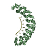
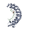
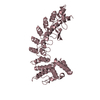
| Title | CRYSTAL STRUCTURE OF THE PUMILIO-HOMOLOGY DOMAIN FROM HUMAN PUMILIO1 IN COMPLEX WITH NRE1-19 RNA | ||||||
 Components Components |
| ||||||
 Keywords Keywords | RNA Binding Protein/RNA / Pumilio-homology domain / Puf domain / Nanos response element / RNA Binding Protein-RNA COMPLEX | ||||||
| Function / homology |  Function and homology information Function and homology informationregulation of miRNA-mediated gene silencing / positive regulation of miRNA-mediated gene silencing /  regulation of chromosome segregation / positive regulation of RIG-I signaling pathway / post-transcriptional gene silencing / 3'-UTR-mediated mRNA destabilization / miRNA processing / miRNA binding / post-transcriptional regulation of gene expression / Golgi Associated Vesicle Biogenesis ...regulation of miRNA-mediated gene silencing / positive regulation of miRNA-mediated gene silencing / regulation of chromosome segregation / positive regulation of RIG-I signaling pathway / post-transcriptional gene silencing / 3'-UTR-mediated mRNA destabilization / miRNA processing / miRNA binding / post-transcriptional regulation of gene expression / Golgi Associated Vesicle Biogenesis / mRNA destabilization / regulation of mRNA stability / adult locomotory behavior / mRNA 3'-UTR binding / stem cell differentiation / P-body / cytoplasmic stress granule / regulation of translation / spermatogenesis / regulation of cell cycle / axon / RNA binding / nucleoplasm / cytosol / cytoplasm regulation of chromosome segregation / positive regulation of RIG-I signaling pathway / post-transcriptional gene silencing / 3'-UTR-mediated mRNA destabilization / miRNA processing / miRNA binding / post-transcriptional regulation of gene expression / Golgi Associated Vesicle Biogenesis ...regulation of miRNA-mediated gene silencing / positive regulation of miRNA-mediated gene silencing / regulation of chromosome segregation / positive regulation of RIG-I signaling pathway / post-transcriptional gene silencing / 3'-UTR-mediated mRNA destabilization / miRNA processing / miRNA binding / post-transcriptional regulation of gene expression / Golgi Associated Vesicle Biogenesis / mRNA destabilization / regulation of mRNA stability / adult locomotory behavior / mRNA 3'-UTR binding / stem cell differentiation / P-body / cytoplasmic stress granule / regulation of translation / spermatogenesis / regulation of cell cycle / axon / RNA binding / nucleoplasm / cytosol / cytoplasmSimilarity search - Function | ||||||
| Biological species |  Homo sapiens (human) Homo sapiens (human) | ||||||
| Method | X-RAY DIFFRACTION / SYNCHROTRON / MOLECULAR REPLACEMENT / Resolution: 2.2 Å | ||||||
 Authors Authors | Wang, X. / McLachlan, J. / Zamore, P.D. / Hall, T.M.T. | ||||||
 Citation Citation | Journal: CELL(CAMBRIDGE,MASS.) / Year: 2002 Title: MODULAR RECOGNITION OF RNA BY A HUMAN PUMILIO-HOMOLOGY DOMAIN Authors: Wang, X. / McLachlan, J. / Zamore, P.D. / Hall, T.M.T. #1: Journal: Mol.Cell / Year: 2001Title: Crystal structure of a Pumilio Homology Domain Authors: Wang, X. / Zamore, P.D. / Hall, T.M.T. | ||||||
| History |
| ||||||
| Remark 400 | COMPOUND CHAIN C AND E ARE ALTERNATE CONFORMATIONS OF EACH OTHER. CHAIN D AND F ARE ALTERNATE ...COMPOUND CHAIN C AND E ARE ALTERNATE CONFORMATIONS OF EACH OTHER. CHAIN D AND F ARE ALTERNATE CONFORMATIONS OF EACH OTHER. |
- Structure visualization
| Structure viewer | Molecule: MolmilJmol/JSmol |
|---|
- Downloads & links
-Download
| PDBx/mmCIF format | 1m8w.cif.gz | 168.3 KB | Display | PDBx/mmCIF format |
|---|---|---|---|---|
| PDB format | pdb1m8w.ent.gz | 131.8 KB | Display | PDB format |
| PDBx/mmJSON format | 1m8w.json.gz | Tree view | PDBx/mmJSON format | |
| Others |  Other downloads Other downloads |
-Validation report
| Arichive directory | https://data.pdbj.org/pub/pdb/validation_reports/m8/1m8wftp://data.pdbj.org/pub/pdb/validation_reports/m8/1m8w | HTTPS FTP |
|---|
-Related structure data
| Related structure data |  1m8xC  1m8yC  1ib3 C: citing same article ( S: Starting model for refinement |
|---|---|
| Similar structure data |


-Links
 PDBj
PDBj
- Assembly
| Deposited unit | 
| ||||||||
|---|---|---|---|---|---|---|---|---|---|
| 1 | 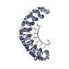
| ||||||||
| 2 | 
| ||||||||
| Unit cell |
| ||||||||
| Details | Two biological units are contained within an asymmetric unit |
-Components
-RNA chain , 4 types, 4 molecules CDEF
| #1: RNA chain | Mass: 2489.489 Da / Num. of mol.: 1 / Source method: obtained synthetically Details: This sequence occurs naturally in Drosophila melanogaster |
|---|---|
| #2: RNA chain | Mass: 2183.323 Da / Num. of mol.: 1 / Source method: obtained synthetically Details: This sequence occurs naturally in Drosophila melanogaster |
| #3: RNA chain | Mass: 2503.521 Da / Num. of mol.: 1 / Source method: obtained synthetically Details: This sequence occurs naturally in Drosophila melanogaster |
| #4: RNA chain | Mass: 2197.355 Da / Num. of mol.: 1 / Source method: obtained synthetically Details: This sequence occurs naturally in Drosophila melanogaster |
-Protein / Non-polymers , 2 types, 309 molecules AB
| #5: Protein | Mass: 40364.523 Da / Num. of mol.: 2 / Fragment: Pumilio-homology domain, Residues 828-1176 Source method: isolated from a genetically manipulated source Source: (gene. exp.) Homo sapiens (human) / Plasmid: pTYB3 / Species (production host): Escherichia coli / Production host:  Escherichia coli BL21(DE3) (bacteria) / Strain (production host): BL21(DE3) / References: UniProt: Q14671 Escherichia coli BL21(DE3) (bacteria) / Strain (production host): BL21(DE3) / References: UniProt: Q14671#6: Water | ChemComp-HOH / | WaterMass: 18.015 Da / Num. of mol.: 307 / Source method: isolated from a natural source / Formula: H2O |
|---|
-Experimental details
-Experiment
| Experiment | Method: X-RAY DIFFRACTION / Number of used crystals: 1 |
|---|
- Sample preparation
| Crystal | Density Matthews: 2.21 Å3/Da / Density % sol: 44.38 % | ||||||||||||||||||||||||||||||||||||||||||||||||||||||||
|---|---|---|---|---|---|---|---|---|---|---|---|---|---|---|---|---|---|---|---|---|---|---|---|---|---|---|---|---|---|---|---|---|---|---|---|---|---|---|---|---|---|---|---|---|---|---|---|---|---|---|---|---|---|---|---|---|---|
| Crystal grow | Temperature: 293 K / Method: vapor diffusion, hanging drop / pH: 5.6 Details: PEG 3350, lithium sulfate, sodium citrate, pH 5.6, VAPOR DIFFUSION, HANGING DROP, temperature 293K | ||||||||||||||||||||||||||||||||||||||||||||||||||||||||
| Crystal grow | *PLUS Temperature: 20 ℃ / pH: 7.4 | ||||||||||||||||||||||||||||||||||||||||||||||||||||||||
| Components of the solutions | *PLUS
|
-Data collection
| Diffraction | Mean temperature: 93 K |
|---|---|
| Diffraction source | Source: SYNCHROTRON / Site: NSLS  / Beamline: X9B / Wavelength: 1.0093 Å / Beamline: X9B / Wavelength: 1.0093 Å |
| Detector | Type: ADSC QUANTUM 4 / Detector: CCD / Date: Mar 7, 2001 |
| Radiation | Protocol: SINGLE WAVELENGTH / Monochromatic (M) / Laue (L): M / Scattering type: x-ray |
| Radiation wavelength | Wavelength: 1.0093 Å / Relative weight: 1 |
| Reflection | Resolution: 2.2→42.68 Å / Num. all: 40790 / Num. obs: 40790 / % possible obs: 99.3 % / Observed criterion σ(I): -3 / Redundancy: 3.1 % / Biso Wilson estimate: 25.7 Å2 / Rsym value: 0.042 / Net I/σ(I): 25.9 |
| Reflection shell | Resolution: 2.2→2.28 Å / Redundancy: 2.8 % / Mean I/σ(I) obs: 3 / Num. unique all: 3936 / Rsym value: 0.359 / % possible all: 97.8 |
| Reflection | *PLUS Rmerge(I) obs: 0.042 |
| Reflection shell | *PLUS % possible obs: 97.8 % / Num. unique obs: 3936 / Rmerge(I) obs: 0.359 |
- Processing
| Software |
| ||||||||||||||||||||||||||||||||||||
|---|---|---|---|---|---|---|---|---|---|---|---|---|---|---|---|---|---|---|---|---|---|---|---|---|---|---|---|---|---|---|---|---|---|---|---|---|---|
| Refinement | Method to determine structure: MOLECULAR REPLACEMENT Starting model: PDB ENTRY 1IB3 1ib3 Resolution: 2.2→42.68 Å / Rfactor Rfree error: 0.006 / Isotropic thermal model: RESTRAINED / Cross valid method: THROUGHOUT / σ(F): 0 / Stereochemistry target values: Engh & Huber / Details: BULK SOLVENT MODEL USED
| ||||||||||||||||||||||||||||||||||||
| Solvent computation | Solvent model: FLAT MODEL / Bsol: 44.8275 Å2 / ksol: 0.320912 e/Å3 | ||||||||||||||||||||||||||||||||||||
| Displacement parameters | Biso mean: 47.4 Å2
| ||||||||||||||||||||||||||||||||||||
| Refine analyze | Luzzati coordinate error free: 0.38 Å / Luzzati sigma a free: 0.3 Å | ||||||||||||||||||||||||||||||||||||
| Refinement step | Cycle: LAST / Resolution: 2.2→42.68 Å
| ||||||||||||||||||||||||||||||||||||
| Refine LS restraints |
| ||||||||||||||||||||||||||||||||||||
| LS refinement shell | Resolution: 2.2→2.34 Å / Rfactor Rfree error: 0.017 / Total num. of bins used: 6
| ||||||||||||||||||||||||||||||||||||
| Xplor file |
| ||||||||||||||||||||||||||||||||||||
| Refinement | *PLUS Highest resolution: 2.2 Å / % reflection Rfree: 6 % / Rfactor all: 0.2279 / Rfactor obs: 0.225 / Rfactor Rfree: 0.29 / Rfactor Rwork: 0.224 | ||||||||||||||||||||||||||||||||||||
| Solvent computation | *PLUS | ||||||||||||||||||||||||||||||||||||
| Displacement parameters | *PLUS | ||||||||||||||||||||||||||||||||||||
| Refine LS restraints | *PLUS
| ||||||||||||||||||||||||||||||||||||
| LS refinement shell | *PLUS Rfactor Rfree: 0.323 / Rfactor Rwork: 0.278 |
