 Movie
Movie Controller


+ Open data

- Basic information
| Entry | Database: PDB / ID: 1m5p | ||||||
|---|---|---|---|---|---|---|---|
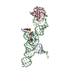
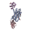
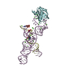
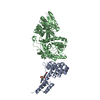
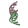
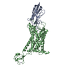
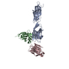
| Title | Transition State Stabilization by a Catalytic RNA | ||||||
 Components Components |
| ||||||
 Keywords Keywords | TRANSLATION/RNA /  HAIRPIN RIBOZYME / CATALYTIC RNA / U1A RNA BINDING PROTEIN / CL5* / TRANSLATION-RNA COMPLEX HAIRPIN RIBOZYME / CATALYTIC RNA / U1A RNA BINDING PROTEIN / CL5* / TRANSLATION-RNA COMPLEX | ||||||
| Function / homology |  Function and homology information Function and homology informationU1 snRNP binding / U1 snRNP / U1 snRNA binding / U4/U6 x U5 tri-snRNP complex / mRNA Splicing - Major Pathway / spliceosomal complex / mRNA splicing, via spliceosome / DNA binding / RNA binding / nucleoplasm ...U1 snRNP binding / U1 snRNP / U1 snRNA binding / U4/U6 x U5 tri-snRNP complex / mRNA Splicing - Major Pathway / spliceosomal complex / mRNA splicing, via spliceosome / DNA binding / RNA binding / nucleoplasm / identical protein binding / nucleusSimilarity search - Function | ||||||
| Biological species |  Homo sapiens (human) Homo sapiens (human) | ||||||
| Method | X-RAY DIFFRACTION / MOLECULAR REPLACEMENT / Resolution: 2.6 Å | ||||||
 Authors Authors | Rupert, P.B. / Massey, A. / Sigurdsson, S.T. / Ferre-D'Amare, A.R. | ||||||
 Citation Citation | Journal: Science / Year: 2002 Title: Transition state stabilization by a catalytic RNA Authors: Rupert, P.B. / Massey, A.P. / Sigurdsson, S.T. / Ferre-D'Amare, A.R. #1: Journal: Nature / Year: 2001Title: Crystal structure of a hairpin ribozyme-inhibitor complex with implications for catalysis Authors: Rupert, P.B. / Ferre-D'Amare, A.R. | ||||||
| History |
|
- Structure visualization
| Structure viewer | Molecule: MolmilJmol/JSmol |
|---|
- Downloads & links
-Download
| PDBx/mmCIF format | 1m5p.cif.gz | 176.8 KB | Display | PDBx/mmCIF format |
|---|---|---|---|---|
| PDB format | pdb1m5p.ent.gz | 132.6 KB | Display | PDB format |
| PDBx/mmJSON format | 1m5p.json.gz | Tree view | PDBx/mmJSON format | |
| Others |  Other downloads Other downloads |
-Validation report
| Arichive directory | https://data.pdbj.org/pub/pdb/validation_reports/m5/1m5pftp://data.pdbj.org/pub/pdb/validation_reports/m5/1m5p | HTTPS FTP |
|---|
-Related structure data
| Related structure data | 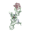 1m5oC  1m5vC  1hp6 C: citing same article ( S: Starting model for refinement |
|---|---|
| Similar structure data |


-Links
 PDBj
PDBj


- Assembly
| Deposited unit | 
| ||||||||||
|---|---|---|---|---|---|---|---|---|---|---|---|
| 1 | 
| ||||||||||
| 2 | 
| ||||||||||
| Unit cell |
|
-Components
-RNA INHIBITOR ... , 2 types, 4 molecules ADMP
| #1: RNA chain | Mass: 3810.352 Da / Num. of mol.: 2 / Source method: obtained synthetically Details: THIS SEQUENCE OCCURS NATURALLY IN SATELLITE TOBACCO RINGSPOT VIRUS #2: RNA chain | Mass: 2683.120 Da / Num. of mol.: 2 / Source method: obtained synthetically Details: THIS SEQUENCE OCCURS NATURALLY IN SATELLITE TOBACCO RINGSPOT VIRUS |
|---|
-RNA chain / Protein , 2 types, 4 molecules BECF
| #3: RNA chain | Mass: 29809.795 Da / Num. of mol.: 2 / Source method: obtained synthetically Details: THIS SEQUENCE OCCURS NATURALLY IN SATELLITE TOBACCO RINGSPOT VIRUS #4: Protein | Mass: 11498.472 Da / Num. of mol.: 2 / Fragment: U1A RNA BINDING DOMAIN / Mutation: Y31H,Q36R Source method: isolated from a genetically manipulated source Source: (gene. exp.) Homo sapiens (human) / Gene: SNRPA / Production host:  Escherichia coli (E. coli) / References: UniProt: P09012 Escherichia coli (E. coli) / References: UniProt: P09012 |
|---|
-Non-polymers , 2 types, 71 molecules 

| #5: Chemical | ChemComp-CA /  Mass: 40.078 Da / Num. of mol.: 26 / Source method: obtained synthetically / Formula: Ca Mass: 40.078 Da / Num. of mol.: 26 / Source method: obtained synthetically / Formula: Ca#6: Water | ChemComp-HOH / | WaterMass: 18.015 Da / Num. of mol.: 45 / Source method: isolated from a natural source / Formula: H2O |
|---|
-Experimental details
-Experiment
| Experiment | Method: X-RAY DIFFRACTION / Number of used crystals: 1 |
|---|
- Sample preparation
| Crystal | Density Matthews: 2.95 Å3/Da / Density % sol: 58.26 % | ||||||||||||||||||||||||||||||||
|---|---|---|---|---|---|---|---|---|---|---|---|---|---|---|---|---|---|---|---|---|---|---|---|---|---|---|---|---|---|---|---|---|---|
| Crystal grow | Temperature: 300 K / Method: vapor diffusion, sitting drop / pH: 5 Details: MPD, calcium Chloride, ammonium chloride, meta-ammonium vanadate, pH 5.0, VAPOR DIFFUSION, SITTING DROP at 300K | ||||||||||||||||||||||||||||||||
| Components of the solutions |
|
-Data collection
| Diffraction | Mean temperature: 150 K |
|---|---|
| Diffraction source | Source: ROTATING ANODE / Type: RIGAKU RU200 / Wavelength: 1.5418 Å |
| Detector | Type: RIGAKU RAXIS IV / Detector: IMAGE PLATE / Date: Dec 12, 2001 / Details: Osmic mirrors |
| Radiation | Monochromator: osmic mirrors / Protocol: SINGLE WAVELENGTH / Monochromatic (M) / Laue (L): M / Scattering type: x-ray |
| Radiation wavelength | Wavelength: 1.5418 Å / Relative weight: 1 |
| Reflection | Resolution: 2.6→30 Å / Num. all: 34446 / Num. obs: 34446 / % possible obs: 98.8 % / Observed criterion σ(F): 0 / Observed criterion σ(I): 0 / Redundancy: 3.3 % / Biso Wilson estimate: 70.9 Å2 / Rsym value: 0.066 / Net I/σ(I): 22 |
| Reflection shell | Resolution: 2.6→2.69 Å / Mean I/σ(I) obs: 1.7 / Num. unique all: 3285 / Rsym value: 0.608 / % possible all: 96.4 |
- Processing
| Software |
| ||||||||||||||||||||||||||||||||||||
|---|---|---|---|---|---|---|---|---|---|---|---|---|---|---|---|---|---|---|---|---|---|---|---|---|---|---|---|---|---|---|---|---|---|---|---|---|---|
| Refinement | Method to determine structure: MOLECULAR REPLACEMENT Starting model: PDB entry 1HP6 1hp6 Resolution: 2.6→27.7 Å / Rfactor Rfree error: 0.005 / Isotropic thermal model: RESTRAINED / Cross valid method: THROUGHOUT / σ(F): 0 / σ(I): 0 Stereochemistry target values: Engh & Huber, Parkinson et al.
| ||||||||||||||||||||||||||||||||||||
| Solvent computation | Solvent model: FLAT MODEL / Bsol: 45.1814 Å2 / ksol: 0.273454 e/Å3 | ||||||||||||||||||||||||||||||||||||
| Displacement parameters | Biso mean: 93.7 Å2
| ||||||||||||||||||||||||||||||||||||
| Refine analyze | Luzzati coordinate error free: 0.54 Å / Luzzati sigma a free: 0.63 Å | ||||||||||||||||||||||||||||||||||||
| Refinement step | Cycle: LAST / Resolution: 2.6→27.7 Å
| ||||||||||||||||||||||||||||||||||||
| Refine LS restraints |
| ||||||||||||||||||||||||||||||||||||
| LS refinement shell | Resolution: 2.6→2.76 Å / Rfactor Rfree error: 0.021 / Total num. of bins used: 6
| ||||||||||||||||||||||||||||||||||||
| Xplor file |
|
