 Movie
Movie Controller

[English] 日本語
 Yorodumi
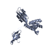
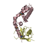
- PDB-1m1h: Crystal structure of Aquifex aeolicus N-utilization substance G (... -
+ Open data

- Basic information
| Entry | Database: PDB / ID: 1m1h | ||||||
|---|---|---|---|---|---|---|---|
| Title | Crystal structure of Aquifex aeolicus N-utilization substance G (NusG), Space group I222 | ||||||
 Components Components | Transcription antitermination protein nusG | ||||||
 Keywords Keywords |  TRANSCRIPTION / Transcription termination / Antitermination / RNP motif / immunoglobulin fold / nucleic acid interaction / protein-protein interaction TRANSCRIPTION / Transcription termination / Antitermination / RNP motif / immunoglobulin fold / nucleic acid interaction / protein-protein interaction | ||||||
| Function / homology |  Function and homology information Function and homology informationtranscription elongation-coupled chromatin remodeling / regulation of DNA-templated transcription elongation / transcription antitermination / DNA-templated transcription termination / cytosolSimilarity search - Function | ||||||
| Biological species |   Aquifex aeolicus (bacteria) Aquifex aeolicus (bacteria) | ||||||
| Method | X-RAY DIFFRACTION / SYNCHROTRON / MIR / Resolution: 1.95 Å | ||||||
 Authors Authors | Steiner, T. / Kaiser, J.T. / Marinkovic, S. / Huber, R. / Wahl, M.C. | ||||||
 Citation Citation | Journal: Embo J. / Year: 2002 Title: Crystal structures of transcription factor NusG in light of its nucleic acid- and protein-binding activities Authors: Steiner, T. / Kaiser, J.T. / Marinkovic, S. / Huber, R. / Wahl, M.C. | ||||||
| History |
|
- Structure visualization
| Structure viewer | Molecule: MolmilJmol/JSmol |
|---|
- Downloads & links
-Download
| PDBx/mmCIF format | 1m1h.cif.gz | 52.6 KB | Display | PDBx/mmCIF format |
|---|---|---|---|---|
| PDB format | pdb1m1h.ent.gz | 41 KB | Display | PDB format |
| PDBx/mmJSON format | 1m1h.json.gz | Tree view | PDBx/mmJSON format | |
| Others |  Other downloads Other downloads |
-Validation report
| Arichive directory | https://data.pdbj.org/pub/pdb/validation_reports/m1/1m1hftp://data.pdbj.org/pub/pdb/validation_reports/m1/1m1h | HTTPS FTP |
|---|
-Related structure data


-Links
 PDBj
PDBj

- Assembly
| Deposited unit | 
| ||||||||
|---|---|---|---|---|---|---|---|---|---|
| 1 |
| ||||||||
| Unit cell |
| ||||||||
| Details | The biological assembly is a monomer |
-Components
| #1: Protein | Mass: 28041.703 Da / Num. of mol.: 1 Source method: isolated from a genetically manipulated source Source: (gene. exp.) Aquifex aeolicus (bacteria) / Production host: Escherichia coli (E. coli) / References: UniProt: O67757 |
|---|---|
| #2: Water | ChemComp-HOH / Water Mass: 18.015 Da / Num. of mol.: 296 / Source method: isolated from a natural source / Formula: H2O Mass: 18.015 Da / Num. of mol.: 296 / Source method: isolated from a natural source / Formula: H2O |
-Experimental details
-Experiment
| Experiment | Method: X-RAY DIFFRACTION / Number of used crystals: 1 |
|---|
- Sample preparation
| Crystal | Density Matthews: 3.38 Å3/Da / Density % sol: 63.57 % | ||||||||||||||||||||||||||||||||||||||||||||||||||||||||
|---|---|---|---|---|---|---|---|---|---|---|---|---|---|---|---|---|---|---|---|---|---|---|---|---|---|---|---|---|---|---|---|---|---|---|---|---|---|---|---|---|---|---|---|---|---|---|---|---|---|---|---|---|---|---|---|---|---|
| Crystal grow | Temperature: 291 K / Method: vapor diffusion, sitting drop / pH: 5.8 Details: PEG 4000, 2-Propanol, Sodium Chloride, pH 5.8, VAPOR DIFFUSION, SITTING DROP, temperature 291K | ||||||||||||||||||||||||||||||||||||||||||||||||||||||||
| Crystal grow | *PLUS Temperature: 18 ℃ / pH: 8.2 | ||||||||||||||||||||||||||||||||||||||||||||||||||||||||
| Components of the solutions | *PLUS
|
-Data collection
| Diffraction | Mean temperature: 100 K |
|---|---|
| Diffraction source | Source: SYNCHROTRON / Site: MPG/DESY, HAMBURG  / Beamline: BW6 / Wavelength: 0.95 Å / Beamline: BW6 / Wavelength: 0.95 Å |
| Detector | Type: MARRESEARCH / Detector: CCD / Details: mirrors |
| Radiation | Monochromator: SAGITALLY FOCUSED Si(111) / Protocol: SINGLE WAVELENGTH / Monochromatic (M) / Laue (L): M / Scattering type: x-ray |
| Radiation wavelength | Wavelength: 0.95 Å / Relative weight: 1 |
| Reflection | Resolution: 1.95→30 Å / Num. all: 27602 / Num. obs: 27602 / % possible obs: 98.5 % / Observed criterion σ(F): 1 / Observed criterion σ(I): 1 / Redundancy: 3.6 % / Rsym value: 0.074 / Net I/σ(I): 30 |
| Reflection shell | Resolution: 1.95→2.05 Å / Mean I/σ(I) obs: 2.3 / Rsym value: 0.504 / % possible all: 98.4 |
| Reflection | *PLUS Rmerge(I) obs: 0.074 |
| Reflection shell | *PLUS % possible obs: 98.4 % / Rmerge(I) obs: 0.504 |
- Processing
| Software |
| |||||||||||||||||||||||||
|---|---|---|---|---|---|---|---|---|---|---|---|---|---|---|---|---|---|---|---|---|---|---|---|---|---|---|
| Refinement | Method to determine structure: MIR / Resolution: 1.95→30 Å / Isotropic thermal model: Isotropic / Cross valid method: THROUGHOUT / σ(F): 0 / σ(I): 0 / Stereochemistry target values: Engh & Huber
| |||||||||||||||||||||||||
| Refinement step | Cycle: LAST / Resolution: 1.95→30 Å
| |||||||||||||||||||||||||
| Refine LS restraints |
| |||||||||||||||||||||||||
| Refinement | *PLUS | |||||||||||||||||||||||||
| Solvent computation | *PLUS | |||||||||||||||||||||||||
| Displacement parameters | *PLUS | |||||||||||||||||||||||||
| LS refinement shell | *PLUS Rfactor Rfree: 0.286 / Rfactor Rwork: 0.283 |
