 Movie
Movie Controller

[English] 日本語
 Yorodumi
- PDB-1m0q: Structure of Dialkylglycine Decarboxylase Complexed with S-1-amin... -
+ Open data

- Basic information
| Entry | Database: PDB / ID: 1m0q | ||||||
|---|---|---|---|---|---|---|---|
| Title | Structure of Dialkylglycine Decarboxylase Complexed with S-1-aminoethanephosphonate | ||||||
 Components Components | 2,2-Dialkylglycine Decarboxylase | ||||||
 Keywords Keywords |  LYASE / Decarboxylase / Pyridoxal phosphate LYASE / Decarboxylase / Pyridoxal phosphate | ||||||
| Function / homology |  Function and homology information2,2-dialkylglycine decarboxylase (pyruvate) / 2,2-dialkylglycine decarboxylase (pyruvate) activity / L-alanine catabolic process, by transamination / alanine-glyoxylate transaminase activity / glyoxylate catabolic process / pyridoxal phosphate binding Function and homology information2,2-dialkylglycine decarboxylase (pyruvate) / 2,2-dialkylglycine decarboxylase (pyruvate) activity / L-alanine catabolic process, by transamination / alanine-glyoxylate transaminase activity / glyoxylate catabolic process / pyridoxal phosphate bindingSimilarity search - Function | ||||||
| Biological species |  Burkholderia cepacia (bacteria) Burkholderia cepacia (bacteria) | ||||||
| Method | X-RAY DIFFRACTION / SYNCHROTRON / FOURIER SYNTHESIS / Resolution: 2 Å | ||||||
 Authors Authors | Liu, W. / Rogers, C.J. / Fisher, A.J. / Toney, M.D. | ||||||
 Citation Citation | Journal: Biochemistry / Year: 2002 Title: Aminophosphonate Inhibitors of Dialkylglycine Decarboxylase: Structural Basis for Slow Binding Inhibition Authors: Liu, W. / Rogers, C.J. / Fisher, A.J. / Toney, M.D. | ||||||
| History |
| ||||||
| Remark 999 | SEQUENCE AUTHORS STATE THAT RESIDUES GLN 15 AND GLY 81 FROM THE SWISSPROT ENTRY ARE INCORRECT. ...SEQUENCE AUTHORS STATE THAT RESIDUES GLN 15 AND GLY 81 FROM THE SWISSPROT ENTRY ARE INCORRECT. THESE RESIDUES SHOULD BE HIS 15 AND GLU 81 AS PRESENT IN AUTHORS' SEQUENCE. |
- Structure visualization
| Structure viewer | Molecule: MolmilJmol/JSmol |
|---|
- Downloads & links
-Download
| PDBx/mmCIF format | 1m0q.cif.gz | 101.1 KB | Display | PDBx/mmCIF format |
|---|---|---|---|---|
| PDB format | pdb1m0q.ent.gz | 76.1 KB | Display | PDB format |
| PDBx/mmJSON format | 1m0q.json.gz | Tree view | PDBx/mmJSON format | |
| Others |  Other downloads Other downloads |
-Validation report
| Arichive directory | https://data.pdbj.org/pub/pdb/validation_reports/m0/1m0qftp://data.pdbj.org/pub/pdb/validation_reports/m0/1m0q | HTTPS FTP |
|---|
-Related structure data
| Related structure data |  1m0nC  1m0oC  1m0pC 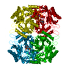 1dkaS C: citing same article ( S: Starting model for refinement |
|---|---|
| Similar structure data |


-Links
 PDBj
PDBj- Assembly
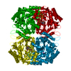
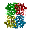
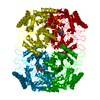
| Deposited unit | 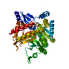
| ||||||||
|---|---|---|---|---|---|---|---|---|---|
| 1 | 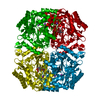
| ||||||||
| Unit cell |
|
-Components
| #1: Protein | Mass: 46577.402 Da / Num. of mol.: 1 Source method: isolated from a genetically manipulated source Source: (gene. exp.) Burkholderia cepacia (bacteria) / Plasmid: pBTac1 / Production host: Escherichia coli (E. coli)References: UniProt: P16932, 2,2-dialkylglycine decarboxylase (pyruvate) |
|---|---|
| #2: Chemical | ChemComp-K /   Mass: 39.098 Da / Num. of mol.: 1 / Source method: obtained synthetically / Formula: K Mass: 39.098 Da / Num. of mol.: 1 / Source method: obtained synthetically / Formula: K |
| #3: Chemical | ChemComp-NA /   Mass: 22.990 Da / Num. of mol.: 1 / Source method: obtained synthetically / Formula: Na Mass: 22.990 Da / Num. of mol.: 1 / Source method: obtained synthetically / Formula: Na |
| #4: Chemical | ChemComp-EPC / (  Mass: 354.190 Da / Num. of mol.: 1 / Source method: obtained synthetically / Formula: C10H16N2O8P2 Mass: 354.190 Da / Num. of mol.: 1 / Source method: obtained synthetically / Formula: C10H16N2O8P2 |
| #5: Water | ChemComp-HOH / Water Mass: 18.015 Da / Num. of mol.: 235 / Source method: isolated from a natural source / Formula: H2O Mass: 18.015 Da / Num. of mol.: 235 / Source method: isolated from a natural source / Formula: H2O |
-Experimental details
-Experiment
| Experiment | Method: X-RAY DIFFRACTION / Number of used crystals: 1 |
|---|
- Sample preparation
| Crystal | Density Matthews: 3 Å3/Da / Density % sol: 61 % | ||||||||||||||||||||||||||||||||||||||||||||||||||||||
|---|---|---|---|---|---|---|---|---|---|---|---|---|---|---|---|---|---|---|---|---|---|---|---|---|---|---|---|---|---|---|---|---|---|---|---|---|---|---|---|---|---|---|---|---|---|---|---|---|---|---|---|---|---|---|---|
| Crystal grow | pH: 6.4 Details: 15% PEG 4000, 0.20 - 0.40 M SODIUM PYRUVATE, 0.015 M MES-KOH (pH 6.4) | ||||||||||||||||||||||||||||||||||||||||||||||||||||||
| Crystal grow | *PLUS pH: 7.5 / Method: vapor diffusion, hanging drop | ||||||||||||||||||||||||||||||||||||||||||||||||||||||
| Components of the solutions | *PLUS
|
-Data collection
| Diffraction | Mean temperature: 100 K |
|---|---|
| Diffraction source | Source: SYNCHROTRON / Site: SSRL  / Beamline: BL9-1 / Wavelength: 0.98 Å / Beamline: BL9-1 / Wavelength: 0.98 Å |
| Detector | Type: MARRESEARCH / Detector: CCD / Date: Feb 28, 2002 |
| Radiation | Protocol: SINGLE WAVELENGTH / Monochromatic (M) / Laue (L): M / Scattering type: x-ray |
| Radiation wavelength | Wavelength: 0.98 Å / Relative weight: 1 |
| Reflection | Resolution: 2→30 Å / Num. all: 38642 / Num. obs: 38642 / % possible obs: 99.2 % / Observed criterion σ(F): 2 / Observed criterion σ(I): -2 / Redundancy: 6.57 % / Rmerge(I) obs: 0.059 / Net I/σ(I): 13.4 |
| Reflection shell | Resolution: 2→2.05 Å / % possible obs: 97.3 % / Redundancy: 6.2 % / Rmerge(I) obs: 0.407 / Mean I/σ(I) obs: 4.2 / % possible all: 99.2 |
| Reflection | *PLUS Lowest resolution: 20 Å / Rmerge(I) obs: 0.059 |
| Reflection shell | *PLUS Rmerge(I) obs: 0.407 |
- Processing
| Software |
| ||||||||||||||||||||||||||||||
|---|---|---|---|---|---|---|---|---|---|---|---|---|---|---|---|---|---|---|---|---|---|---|---|---|---|---|---|---|---|---|---|
| Refinement | Method to determine structure: FOURIER SYNTHESIS Starting model: PDB ENTRY 1DKA Resolution: 2→20 Å / Isotropic thermal model: restrained / Cross valid method: THROUGHOUT / σ(F): 2 / Stereochemistry target values: Engh & Huber
| ||||||||||||||||||||||||||||||
| Solvent computation | Bsol: 157 Å2 / ksol: 0.78 e/Å3 | ||||||||||||||||||||||||||||||
| Refinement step | Cycle: LAST / Resolution: 2→20 Å
| ||||||||||||||||||||||||||||||
| Refine LS restraints |
| ||||||||||||||||||||||||||||||
| Refinement | *PLUS Lowest resolution: 20 Å / % reflection Rfree: 5 % / Rfactor obs: 0.194 / Rfactor Rfree: 0.24 / Rfactor Rwork: 0.194 | ||||||||||||||||||||||||||||||
| Solvent computation | *PLUS | ||||||||||||||||||||||||||||||
| Displacement parameters | *PLUS | ||||||||||||||||||||||||||||||
| Refine LS restraints | *PLUS
|
