 Movie
Movie Controller

[English] 日本語
 Yorodumi
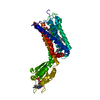
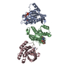
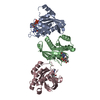
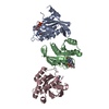
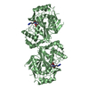
- PDB-1lx5: Crystal Structure of the BMP7/ActRII Extracellular Domain Complex -
+ Open data

- Basic information
| Entry | Database: PDB / ID: 1lx5 | |||||||||
|---|---|---|---|---|---|---|---|---|---|---|
| Title | Crystal Structure of the BMP7/ActRII Extracellular Domain Complex | |||||||||
 Components Components |
| |||||||||
 Keywords Keywords | GROWTH FACTOR/GROWTH FACTOR RECEPTOR / LIGAND-RECEPTOR COMPLEX / GROWTH FACTOR-GROWTH FACTOR RECEPTOR COMPLEX | |||||||||
| Function / homology |  Function and homology information Function and homology informationnegative regulation of prostatic bud formation / negative regulation of mesenchymal cell apoptotic process involved in nephron morphogenesis / negative regulation of glomerular mesangial cell proliferation / mesenchymal cell apoptotic process involved in nephron morphogenesis / positive regulation of cardiac neural crest cell migration involved in outflow tract morphogenesis / positive regulation of hyaluranon cable assembly / Signaling by Activin / chorio-allantoic fusion / inhibin-betaglycan-ActRII complex / negative regulation of striated muscle cell apoptotic process ...negative regulation of prostatic bud formation / negative regulation of mesenchymal cell apoptotic process involved in nephron morphogenesis / negative regulation of glomerular mesangial cell proliferation / mesenchymal cell apoptotic process involved in nephron morphogenesis / positive regulation of cardiac neural crest cell migration involved in outflow tract morphogenesis / positive regulation of hyaluranon cable assembly / Signaling by Activin / chorio-allantoic fusion / inhibin-betaglycan-ActRII complex / negative regulation of striated muscle cell apoptotic process / metanephric mesenchyme morphogenesis / nephrogenic mesenchyme morphogenesis /  inhibin binding / Signaling by BMP / embryonic skeletal joint morphogenesis / penile erection / neural fold elevation formation / ameloblast differentiation / positive regulation of activin receptor signaling pathway / activin receptor activity / positive regulation of epithelial cell differentiation / metanephric mesenchymal cell proliferation involved in metanephros development / embryonic camera-type eye morphogenesis / mesenchyme development / positive regulation of follicle-stimulating hormone secretion / cellular response to oxygen-glucose deprivation / Sertoli cell proliferation / sperm ejaculation / allantois development / mesonephros development / BMP receptor activity / embryonic skeletal system development / monocyte aggregation / regulation of removal of superoxide radicals / hindbrain development / BMP receptor binding / activin receptor complex / mesenchymal cell differentiation / pericardium morphogenesis / endocardial cushion formation / receptor protein serine/threonine kinase / pharyngeal system development / heart trabecula morphogenesis / activin binding / cellular response to BMP stimulus / branching involved in salivary gland morphogenesis / embryonic pattern specification / activin receptor signaling pathway / response to vitamin D / regulation of phosphorylation / SMAD protein signal transduction / metanephros development / cardiac muscle tissue development / negative regulation of mitotic nuclear division / gastrulation with mouth forming second / regulation of nitric oxide biosynthetic process / negative regulation of non-canonical NF-kappaB signal transduction / cartilage development / positive regulation of dendrite development / determination of left/right symmetry / embryonic limb morphogenesis / Molecules associated with elastic fibres / positive regulation of heterotypic cell-cell adhesion / ureteric bud development / anterior/posterior pattern specification / regulation of branching involved in prostate gland morphogenesis / branching morphogenesis of an epithelial tube / negative regulation of NF-kappaB transcription factor activity / negative regulation of Notch signaling pathway / dendrite development / cardiac septum morphogenesis / odontogenesis of dentin-containing tooth / growth factor binding / mesoderm development / positive regulation of SMAD protein signal transduction / mesoderm formation / negative regulation of cell cycle / negative regulation of neuron differentiation / epithelial to mesenchymal transition / regulation of signal transduction / positive regulation of epithelial to mesenchymal transition / positive regulation of osteoblast differentiation / BMP signaling pathway / positive regulation of bone mineralization / coreceptor activity / positive regulation of neuron differentiation / positive regulation of brown fat cell differentiation / ossification / neuron projection morphogenesis / positive regulation of erythrocyte differentiation / skeletal system development / cytokine activity / PDZ domain binding / axon guidance / growth factor activity / response to peptide hormone / cellular response to growth factor stimulus / autophagy / negative regulation of neurogenesis / male gonad development inhibin binding / Signaling by BMP / embryonic skeletal joint morphogenesis / penile erection / neural fold elevation formation / ameloblast differentiation / positive regulation of activin receptor signaling pathway / activin receptor activity / positive regulation of epithelial cell differentiation / metanephric mesenchymal cell proliferation involved in metanephros development / embryonic camera-type eye morphogenesis / mesenchyme development / positive regulation of follicle-stimulating hormone secretion / cellular response to oxygen-glucose deprivation / Sertoli cell proliferation / sperm ejaculation / allantois development / mesonephros development / BMP receptor activity / embryonic skeletal system development / monocyte aggregation / regulation of removal of superoxide radicals / hindbrain development / BMP receptor binding / activin receptor complex / mesenchymal cell differentiation / pericardium morphogenesis / endocardial cushion formation / receptor protein serine/threonine kinase / pharyngeal system development / heart trabecula morphogenesis / activin binding / cellular response to BMP stimulus / branching involved in salivary gland morphogenesis / embryonic pattern specification / activin receptor signaling pathway / response to vitamin D / regulation of phosphorylation / SMAD protein signal transduction / metanephros development / cardiac muscle tissue development / negative regulation of mitotic nuclear division / gastrulation with mouth forming second / regulation of nitric oxide biosynthetic process / negative regulation of non-canonical NF-kappaB signal transduction / cartilage development / positive regulation of dendrite development / determination of left/right symmetry / embryonic limb morphogenesis / Molecules associated with elastic fibres / positive regulation of heterotypic cell-cell adhesion / ureteric bud development / anterior/posterior pattern specification / regulation of branching involved in prostate gland morphogenesis / branching morphogenesis of an epithelial tube / negative regulation of NF-kappaB transcription factor activity / negative regulation of Notch signaling pathway / dendrite development / cardiac septum morphogenesis / odontogenesis of dentin-containing tooth / growth factor binding / mesoderm development / positive regulation of SMAD protein signal transduction / mesoderm formation / negative regulation of cell cycle / negative regulation of neuron differentiation / epithelial to mesenchymal transition / regulation of signal transduction / positive regulation of epithelial to mesenchymal transition / positive regulation of osteoblast differentiation / BMP signaling pathway / positive regulation of bone mineralization / coreceptor activity / positive regulation of neuron differentiation / positive regulation of brown fat cell differentiation / ossification / neuron projection morphogenesis / positive regulation of erythrocyte differentiation / skeletal system development / cytokine activity / PDZ domain binding / axon guidance / growth factor activity / response to peptide hormone / cellular response to growth factor stimulus / autophagy / negative regulation of neurogenesis / male gonad developmentSimilarity search - Function | |||||||||
| Biological species |  Homo sapiens (human) Homo sapiens (human) Mus musculus (house mouse) Mus musculus (house mouse) | |||||||||
| Method | X-RAY DIFFRACTION / SYNCHROTRON / MIR / Resolution: 3.3 Å | |||||||||
 Authors Authors | Greenwald, J. / Groppe, J. / Kwiatkowski, W. / Choe, S. | |||||||||
 Citation Citation | Journal: Mol.Cell / Year: 2003 Title: The BMP7/ActRII Extracellular Domain Complex Provides New Insights into the Cooperative Nature of Receptor Assembly Authors: Greenwald, J. / Groppe, J. / Gray, P. / Wiater, E. / Kwiatkowski, W. / Vale, W. / Choe, S. | |||||||||
| History |
| |||||||||
| Remark 999 | SEQUENCE THE SEQEUNCE FOR ACVR2 IS TRUNCATED. THE PROTEIN IN THE CRYSTAL ENDS AT GLU 102. |
- Structure visualization
| Structure viewer | Molecule: MolmilJmol/JSmol |
|---|
- Downloads & links
-Download
| PDBx/mmCIF format | 1lx5.cif.gz | 55.2 KB | Display | PDBx/mmCIF format |
|---|---|---|---|---|
| PDB format | pdb1lx5.ent.gz | 43 KB | Display | PDB format |
| PDBx/mmJSON format | 1lx5.json.gz | Tree view | PDBx/mmJSON format | |
| Others |  Other downloads Other downloads |
-Validation report
| Arichive directory | https://data.pdbj.org/pub/pdb/validation_reports/lx/1lx5ftp://data.pdbj.org/pub/pdb/validation_reports/lx/1lx5 | HTTPS FTP |
|---|
-Related structure data


-Links
 PDBj
PDBj


- Assembly
| Deposited unit | 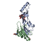
| ||||||||
|---|---|---|---|---|---|---|---|---|---|
| 1 | 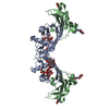
| ||||||||
| Unit cell |
| ||||||||
| Details | The second part of the biological assembly is generated by the two fold axis: y-x+1, y, -z+1 |
-Components
| #1: Protein | Mass: 15699.730 Da / Num. of mol.: 1 Source method: isolated from a genetically manipulated source Source: (gene. exp.) Homo sapiens (human) / Description: DHFR- / Gene: BMP7 / Cell (production host): OVARY CELLS / Production host:  Cricetulus griseus (Chinese hamster) / References: UniProt: P18075 Cricetulus griseus (Chinese hamster) / References: UniProt: P18075 |
|---|---|
| #2: Protein | Mass: 12019.440 Da / Num. of mol.: 1 Fragment: Extracellular Ligand Binding Domain, C-terminal truncation Source method: isolated from a genetically manipulated source Source: (gene. exp.) Mus musculus (house mouse) / Gene: ACVR2 / Production host:  Pichia pastoris (fungus) / Strain (production host): SMD1168 / References: UniProt: P27038 Pichia pastoris (fungus) / Strain (production host): SMD1168 / References: UniProt: P27038 |
| #3: Polysaccharide | alpha-D-mannopyranose-(1-3)-[beta-D-mannopyranose-(1-4)][alpha-D-mannopyranose-(1-6)]beta-D- ...alpha-D-mannopyranose-(1-3)-[beta-D-mannopyranose-(1-4)][alpha-D-mannopyranose-(1-6)]beta-D-mannopyranose-(1-4)-2-acetamido-2-deoxy-beta-D-glucopyranose-(1-4)-2-acetamido-2-deoxy-beta-D-glucopyranose / Mass: 1072.964 Da / Num. of mol.: 1 Source method: isolated from a genetically manipulated source |
| #4: Sugar | N-Acetylglucosamine  Type: D-saccharide, beta linking / Mass: 221.208 Da / Num. of mol.: 2 Type: D-saccharide, beta linking / Mass: 221.208 Da / Num. of mol.: 2Source method: isolated from a genetically manipulated source Formula: C8H15NO6 |
-Experimental details
-Experiment
| Experiment | Method: X-RAY DIFFRACTION / Number of used crystals: 1 |
|---|
- Sample preparation
| Crystal | Density Matthews: 4.69 Å3/Da / Density % sol: 73.79 % | ||||||||||||||||||||||||
|---|---|---|---|---|---|---|---|---|---|---|---|---|---|---|---|---|---|---|---|---|---|---|---|---|---|
| Crystal grow | Temperature: 296 K / Method: vapor diffusion, hanging drop / pH: 7 Details: 1M sodium acetate, 0.1M imidazole, pH 7, VAPOR DIFFUSION, HANGING DROP, temperature 296K | ||||||||||||||||||||||||
| Crystal grow | *PLUS Temperature: 23 ℃ | ||||||||||||||||||||||||
| Components of the solutions | *PLUS
|
-Data collection
| Diffraction | Mean temperature: 100 K |
|---|---|
| Diffraction source | Source: SYNCHROTRON / Site: SSRL  / Beamline: BL11-1 / Wavelength: 1.03 Å / Beamline: BL11-1 / Wavelength: 1.03 Å |
| Detector | Type: ADSC QUANTUM 315 / Detector: CCD / Date: Feb 10, 2002 |
| Radiation | Monochromator: Si 111 / Protocol: SINGLE WAVELENGTH / Monochromatic (M) / Laue (L): M / Scattering type: x-ray |
| Radiation wavelength | Wavelength: 1.03 Å / Relative weight: 1 |
| Reflection | Resolution: 3.3→30 Å / Num. all: 8320 / Num. obs: 8320 / % possible obs: 96.7 % / Observed criterion σ(F): 0 / Observed criterion σ(I): -3 / Biso Wilson estimate: 145 Å2 / Rsym value: 0.055 / Net I/σ(I): 16.5 |
| Reflection shell | Resolution: 3.3→3.42 Å / Rsym value: 0.332 / % possible all: 98.6 |
| Reflection | *PLUS Lowest resolution: 100 Å / Num. measured all: 31531 / Rmerge(I) obs: 0.055 |
| Reflection shell | *PLUS % possible obs: 98.6 % / Rmerge(I) obs: 0.332 |
- Processing
| Software |
| ||||||||||||||||||||||||||||||||||||||||||||||||||||||||||||||||||||||||||||||||||||||||||||||||||||||||||||||||||||||||||||||||||
|---|---|---|---|---|---|---|---|---|---|---|---|---|---|---|---|---|---|---|---|---|---|---|---|---|---|---|---|---|---|---|---|---|---|---|---|---|---|---|---|---|---|---|---|---|---|---|---|---|---|---|---|---|---|---|---|---|---|---|---|---|---|---|---|---|---|---|---|---|---|---|---|---|---|---|---|---|---|---|---|---|---|---|---|---|---|---|---|---|---|---|---|---|---|---|---|---|---|---|---|---|---|---|---|---|---|---|---|---|---|---|---|---|---|---|---|---|---|---|---|---|---|---|---|---|---|---|---|---|---|---|---|
| Refinement | Method to determine structure: MIR / Resolution: 3.3→27.12 Å / Cor.coef. Fo:Fc: 0.899 / Cor.coef. Fo:Fc free: 0.858 / SU B: 8.619 / SU ML: 0.151 / Isotropic thermal model: isotropic / Cross valid method: THROUGHOUT / σ(F): 0 / ESU R: 0.949 / ESU R Free: 0.435 / Stereochemistry target values: Engh & Huber
| ||||||||||||||||||||||||||||||||||||||||||||||||||||||||||||||||||||||||||||||||||||||||||||||||||||||||||||||||||||||||||||||||||
| Solvent computation | Ion probe radii: 0.8 Å / Shrinkage radii: 0.8 Å / VDW probe radii: 1.4 Å | ||||||||||||||||||||||||||||||||||||||||||||||||||||||||||||||||||||||||||||||||||||||||||||||||||||||||||||||||||||||||||||||||||
| Displacement parameters | Biso mean: 69.6 Å2
| ||||||||||||||||||||||||||||||||||||||||||||||||||||||||||||||||||||||||||||||||||||||||||||||||||||||||||||||||||||||||||||||||||
| Refinement step | Cycle: LAST / Resolution: 3.3→27.12 Å
| ||||||||||||||||||||||||||||||||||||||||||||||||||||||||||||||||||||||||||||||||||||||||||||||||||||||||||||||||||||||||||||||||||
| Refine LS restraints |
| ||||||||||||||||||||||||||||||||||||||||||||||||||||||||||||||||||||||||||||||||||||||||||||||||||||||||||||||||||||||||||||||||||
| LS refinement shell | Resolution: 3.3→3.386 Å / Total num. of bins used: 20 /
| ||||||||||||||||||||||||||||||||||||||||||||||||||||||||||||||||||||||||||||||||||||||||||||||||||||||||||||||||||||||||||||||||||
| Refinement TLS params. | Method: refined / Refine-ID: X-RAY DIFFRACTION
| ||||||||||||||||||||||||||||||||||||||||||||||||||||||||||||||||||||||||||||||||||||||||||||||||||||||||||||||||||||||||||||||||||
| Refinement TLS group |
| ||||||||||||||||||||||||||||||||||||||||||||||||||||||||||||||||||||||||||||||||||||||||||||||||||||||||||||||||||||||||||||||||||
| Software | *PLUS Name: REFMAC / Version: 5 / Classification: refinement | ||||||||||||||||||||||||||||||||||||||||||||||||||||||||||||||||||||||||||||||||||||||||||||||||||||||||||||||||||||||||||||||||||
| Refinement | *PLUS Highest resolution: 3.3 Å / Lowest resolution: 100 Å / % reflection Rfree: 5 % | ||||||||||||||||||||||||||||||||||||||||||||||||||||||||||||||||||||||||||||||||||||||||||||||||||||||||||||||||||||||||||||||||||
| Solvent computation | *PLUS | ||||||||||||||||||||||||||||||||||||||||||||||||||||||||||||||||||||||||||||||||||||||||||||||||||||||||||||||||||||||||||||||||||
| Displacement parameters | *PLUS | ||||||||||||||||||||||||||||||||||||||||||||||||||||||||||||||||||||||||||||||||||||||||||||||||||||||||||||||||||||||||||||||||||
| Refine LS restraints | *PLUS
|
