 Movie
Movie Controller

[English] 日本語
 Yorodumi
- PDB-1lr7: Crystal structure of Fs1, the heparin-binding domain of follistat... -
+ Open data

- Basic information
| Entry | Database: PDB / ID: 1lr7 | ||||||
|---|---|---|---|---|---|---|---|
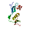
| Title | Crystal structure of Fs1, the heparin-binding domain of follistatin, complexed with the heparin analogue sucrose octasulphate (SOS) | ||||||
 Components Components | follistatin | ||||||
 Keywords Keywords | hormone/growth factor / heparin-binding / cystine-rich / sucrose octasulphate / hormone-growth factor COMPLEX | ||||||
| Function / homology |  Function and homology information Function and homology informationAntagonism of Activin by Follistatin / activin receptor antagonist activity / negative regulation of follicle-stimulating hormone secretion / ameloblast differentiation / positive regulation of hair follicle development / regulation of BMP signaling pathway / gamete generation / pattern specification process / activin binding / negative regulation of activin receptor signaling pathway ...Antagonism of Activin by Follistatin / activin receptor antagonist activity / negative regulation of follicle-stimulating hormone secretion / ameloblast differentiation / positive regulation of hair follicle development / regulation of BMP signaling pathway / gamete generation / pattern specification process / activin binding / negative regulation of activin receptor signaling pathway / heparan sulfate proteoglycan binding / negative regulation of epithelial cell differentiation / hair follicle morphogenesis / female gonad development / odontogenesis of dentin-containing tooth / keratinocyte proliferation / BMP signaling pathway / hematopoietic progenitor cell differentiation / skeletal system development / response to organic cyclic compound / cellular response to growth factor stimulus / cell differentiation / negative regulation of transcription by RNA polymerase II / extracellular space / nucleus / cytoplasmSimilarity search - Function | ||||||
| Biological species |  Rattus norvegicus (Norway rat) Rattus norvegicus (Norway rat) | ||||||
| Method | X-RAY DIFFRACTION / SYNCHROTRON / MAD / Resolution: 1.5 Å | ||||||
 Authors Authors | Innis, C.A. / Hyvonen, M. | ||||||
 Citation Citation | Journal: J.Biol.Chem. / Year: 2003 Title: Crystal Structures of the Heparan Sulfate-binding Domain of Follistatin: Insights into ligand binding. Authors: Innis, C.A. / Hyvonen, M. | ||||||
| History |
| ||||||
| Remark 600 | heterogen Authors claimed that there was a lack of connecting electron density between sulphate ... heterogen Authors claimed that there was a lack of connecting electron density between sulphate groups in the ligand sucrose octasulphate due to possible alternative binding modes. Accordingly, the authors chose to list the sulphate ions as free ions even though they belong to the sucrose octasulphate molecule. |
- Structure visualization
| Structure viewer | Molecule: MolmilJmol/JSmol |
|---|
- Downloads & links
-Download
| PDBx/mmCIF format | 1lr7.cif.gz | 25.3 KB | Display | PDBx/mmCIF format |
|---|---|---|---|---|
| PDB format | pdb1lr7.ent.gz | 18.9 KB | Display | PDB format |
| PDBx/mmJSON format | 1lr7.json.gz | Tree view | PDBx/mmJSON format | |
| Others |  Other downloads Other downloads |
-Validation report
| Arichive directory | https://data.pdbj.org/pub/pdb/validation_reports/lr/1lr7ftp://data.pdbj.org/pub/pdb/validation_reports/lr/1lr7 | HTTPS FTP |
|---|
-Related structure data


-Links
 PDBj
PDBj
- Assembly
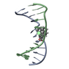
| Deposited unit | 
| ||||||||
|---|---|---|---|---|---|---|---|---|---|
| 1 |
| ||||||||
| Unit cell |
|
-Components
| #1: Protein | / FS1 Mass: 8342.812 Da / Num. of mol.: 1 / Fragment: Heparin-binding domain Source method: isolated from a genetically manipulated source Source: (gene. exp.) Rattus norvegicus (Norway rat) / Plasmid: pBAT4 / Species (production host): Escherichia coli / Production host:  Escherichia coli BL21(DE3) (bacteria) / Strain (production host): BL21(DE3) / References: UniProt: P21674 Escherichia coli BL21(DE3) (bacteria) / Strain (production host): BL21(DE3) / References: UniProt: P21674 | ||
|---|---|---|---|
| #2: Chemical | ChemComp-SO4 / Sulfate  Mass: 96.063 Da / Num. of mol.: 5 / Source method: obtained synthetically / Formula: SO4 Mass: 96.063 Da / Num. of mol.: 5 / Source method: obtained synthetically / Formula: SO4#3: Water | ChemComp-HOH / | Water Mass: 18.015 Da / Num. of mol.: 78 / Source method: isolated from a natural source / Formula: H2O Mass: 18.015 Da / Num. of mol.: 78 / Source method: isolated from a natural source / Formula: H2O |
-Experimental details
-Experiment
| Experiment | Method: X-RAY DIFFRACTION / Number of used crystals: 1 |
|---|
- Sample preparation
| Crystal | Density Matthews: 1.83 Å3/Da / Density % sol: 36.49 % | ||||||||||||||||||||||||||||||
|---|---|---|---|---|---|---|---|---|---|---|---|---|---|---|---|---|---|---|---|---|---|---|---|---|---|---|---|---|---|---|---|
| Crystal grow | Temperature: 289 K / Method: vapor diffusion, hanging drop / pH: 8.5 Details: 30-35% 2-propanol, 0.6-0.7 M ammonium acetate, 0.1 M Tris-HCl, pH 8.5, VAPOR DIFFUSION, HANGING DROP, temperature 289K | ||||||||||||||||||||||||||||||
| Crystal grow | *PLUS Temperature: 18 ℃ / Method: vapor diffusion, hanging drop | ||||||||||||||||||||||||||||||
| Components of the solutions | *PLUS
|
-Data collection
| Diffraction | Mean temperature: 100 K |
|---|---|
| Diffraction source | Source: SYNCHROTRON / Site: ESRF  / Beamline: ID14-4 / Wavelength: 0.9795 Å / Beamline: ID14-4 / Wavelength: 0.9795 Å |
| Detector | Type: ADSC QUANTUM 4 / Detector: CCD / Date: Feb 11, 2001 / Details: Mirrors |
| Radiation | Monochromator: Si 111 CHANNEL / Protocol: SINGLE WAVELENGTH / Monochromatic (M) / Laue (L): M / Scattering type: x-ray |
| Radiation wavelength | Wavelength: 0.9795 Å / Relative weight: 1 |
| Reflection | Resolution: 1.5→39.2 Å / Num. obs: 10456 / % possible obs: 95 % / Observed criterion σ(F): 0 / Observed criterion σ(I): 0 / Redundancy: 5.8 % / Biso Wilson estimate: 14.21 Å2 / Rmerge(I) obs: 0.071 |
| Reflection shell | Resolution: 1.5→1.53 Å / Rmerge(I) obs: 0.196 / % possible all: 71.8 |
| Reflection | *PLUS Rmerge(I) obs: 0.07 |
| Reflection shell | *PLUS % possible obs: 71.8 % / Rmerge(I) obs: 0.2 |
- Processing
| Software |
| ||||||||||||||||||||||||||||||||||||||||||||||||||||||||||||||||||||||||||||||||||||||||||||||||||||||||||||||||||||||||||||||||||
|---|---|---|---|---|---|---|---|---|---|---|---|---|---|---|---|---|---|---|---|---|---|---|---|---|---|---|---|---|---|---|---|---|---|---|---|---|---|---|---|---|---|---|---|---|---|---|---|---|---|---|---|---|---|---|---|---|---|---|---|---|---|---|---|---|---|---|---|---|---|---|---|---|---|---|---|---|---|---|---|---|---|---|---|---|---|---|---|---|---|---|---|---|---|---|---|---|---|---|---|---|---|---|---|---|---|---|---|---|---|---|---|---|---|---|---|---|---|---|---|---|---|---|---|---|---|---|---|---|---|---|---|
| Refinement | Method to determine structure: MAD / Resolution: 1.5→39.22 Å / Cor.coef. Fo:Fc: 0.952 / Cor.coef. Fo:Fc free: 0.932 / SU B: 1.873 / SU ML: 0.07 / Isotropic thermal model: isotropic / Cross valid method: THROUGHOUT / σ(F): 0 / ESU R: 0.086 / ESU R Free: 0.087 / Stereochemistry target values: MAXIMUM LIKELIHOOD / Details: HYDROGENS HAVE BEEN ADDED IN THE RIDING POSITIONS
| ||||||||||||||||||||||||||||||||||||||||||||||||||||||||||||||||||||||||||||||||||||||||||||||||||||||||||||||||||||||||||||||||||
| Solvent computation | Ion probe radii: 0.8 Å / Shrinkage radii: 0.8 Å / VDW probe radii: 1.4 Å / Solvent model: BABINET MODEL WITH MASK | ||||||||||||||||||||||||||||||||||||||||||||||||||||||||||||||||||||||||||||||||||||||||||||||||||||||||||||||||||||||||||||||||||
| Displacement parameters | Biso mean: 15.641 Å2
| ||||||||||||||||||||||||||||||||||||||||||||||||||||||||||||||||||||||||||||||||||||||||||||||||||||||||||||||||||||||||||||||||||
| Refinement step | Cycle: LAST / Resolution: 1.5→39.22 Å
| ||||||||||||||||||||||||||||||||||||||||||||||||||||||||||||||||||||||||||||||||||||||||||||||||||||||||||||||||||||||||||||||||||
| Refine LS restraints |
| ||||||||||||||||||||||||||||||||||||||||||||||||||||||||||||||||||||||||||||||||||||||||||||||||||||||||||||||||||||||||||||||||||
| LS refinement shell | Resolution: 1.5→1.539 Å / Total num. of bins used: 20
| ||||||||||||||||||||||||||||||||||||||||||||||||||||||||||||||||||||||||||||||||||||||||||||||||||||||||||||||||||||||||||||||||||
| Refinement | *PLUS Lowest resolution: 39.2 Å / Rfactor Rfree: 0.222 / Rfactor Rwork: 0.189 | ||||||||||||||||||||||||||||||||||||||||||||||||||||||||||||||||||||||||||||||||||||||||||||||||||||||||||||||||||||||||||||||||||
| Solvent computation | *PLUS | ||||||||||||||||||||||||||||||||||||||||||||||||||||||||||||||||||||||||||||||||||||||||||||||||||||||||||||||||||||||||||||||||||
| Displacement parameters | *PLUS | ||||||||||||||||||||||||||||||||||||||||||||||||||||||||||||||||||||||||||||||||||||||||||||||||||||||||||||||||||||||||||||||||||
| Refine LS restraints | *PLUS
|
