 Movie
Movie Controller

[English] 日本語
 Yorodumi
- PDB-1lr0: Pseudomonas aeruginosa TolA Domain III, Seleno-methionine Derivative -
+ Open data

- Basic information
| Entry | Database: PDB / ID: 1lr0 | ||||||
|---|---|---|---|---|---|---|---|
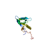
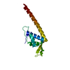
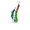
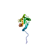
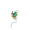
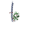
| Title | Pseudomonas aeruginosa TolA Domain III, Seleno-methionine Derivative | ||||||
 Components Components | TolA protein | ||||||
 Keywords Keywords |  PROTEIN TRANSPORT / Domain-Swapping / TolA / TonB PROTEIN TRANSPORT / Domain-Swapping / TolA / TonB | ||||||
| Function / homology |  Function and homology information Function and homology informationbacteriocin transport / toxin transmembrane transporter activity / cell cycle / cell division / plasma membraneSimilarity search - Function | ||||||
| Biological species |   Pseudomonas aeruginosa (bacteria) Pseudomonas aeruginosa (bacteria) | ||||||
| Method | X-RAY DIFFRACTION / SYNCHROTRON / MAD / Resolution: 1.914 Å | ||||||
 Authors Authors | Witty, M. / Sanz, C. / Shah, A. / Grossman, J.G. / Mizuguchi, K. / Perham, R.N. / Luisi, B. | ||||||
 Citation Citation | Journal: EMBO J. / Year: 2002 Title: Structure of the periplasmic domain of Pseudomonas aeruginosa TolA: evidence for an evolutionary relationship with the TonB transporter protein. Authors: Witty, M. / Sanz, C. / Shah, A. / Grossmann, J.G. / Mizuguchi, K. / Perham, R.N. / Luisi, B. | ||||||
| History |
|
- Structure visualization
| Structure viewer | Molecule: MolmilJmol/JSmol |
|---|
- Downloads & links
-Download
| PDBx/mmCIF format | 1lr0.cif.gz | 37.4 KB | Display | PDBx/mmCIF format |
|---|---|---|---|---|
| PDB format | pdb1lr0.ent.gz | 28.8 KB | Display | PDB format |
| PDBx/mmJSON format | 1lr0.json.gz | Tree view | PDBx/mmJSON format | |
| Others |  Other downloads Other downloads |
-Validation report
| Arichive directory | https://data.pdbj.org/pub/pdb/validation_reports/lr/1lr0ftp://data.pdbj.org/pub/pdb/validation_reports/lr/1lr0 | HTTPS FTP |
|---|
-Related structure data
| Similar structure data |
|---|


-Links
 PDBj
PDBj- Assembly
| Deposited unit | 
| ||||||||
|---|---|---|---|---|---|---|---|---|---|
| 1 |
| ||||||||
| 2 | 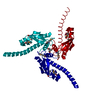
| ||||||||
| Unit cell |
| ||||||||
| Components on special symmetry positions |
|
-Components
| #1: Protein | Mass: 14616.770 Da / Num. of mol.: 1 / Fragment: Periplasmic Domain (residues 226-347) Source method: isolated from a genetically manipulated source Source: (gene. exp.) Pseudomonas aeruginosa (bacteria) / Gene: TOLA / Plasmid: pET-11c / Production host: Escherichia coli (E. coli) / Strain (production host): BL21(DE3)pLysS / References: UniProt: P50600 | ||||
|---|---|---|---|---|---|
| #2: Chemical |   Mass: 65.409 Da / Num. of mol.: 2 / Source method: obtained synthetically / Formula: Zn Mass: 65.409 Da / Num. of mol.: 2 / Source method: obtained synthetically / Formula: Zn#3: Chemical | ChemComp-TRS / | Tris  Mass: 122.143 Da / Num. of mol.: 1 / Source method: obtained synthetically / Formula: C4H12NO3 / Comment: pH buffer*YM Mass: 122.143 Da / Num. of mol.: 1 / Source method: obtained synthetically / Formula: C4H12NO3 / Comment: pH buffer*YM#4: Water | ChemComp-HOH / | Water Mass: 18.015 Da / Num. of mol.: 122 / Source method: isolated from a natural source / Formula: H2O Mass: 18.015 Da / Num. of mol.: 122 / Source method: isolated from a natural source / Formula: H2O |
-Experimental details
-Experiment
| Experiment | Method: X-RAY DIFFRACTION / Number of used crystals: 1 |
|---|
- Sample preparation
| Crystal | Density Matthews: 2.97 Å3/Da / Density % sol: 58.57 % | |||||||||||||||||||||||||||||||||||||||||||||||||
|---|---|---|---|---|---|---|---|---|---|---|---|---|---|---|---|---|---|---|---|---|---|---|---|---|---|---|---|---|---|---|---|---|---|---|---|---|---|---|---|---|---|---|---|---|---|---|---|---|---|---|
| Crystal grow | Temperature: 293 K / Method: vapor diffusion, hanging drop / pH: 8 Details: 5% wt/v PEG 6000, 100 mM TrisCl, pH 8.0, and 1 mM ZnSO4, VAPOR DIFFUSION, HANGING DROP, temperature 293K | |||||||||||||||||||||||||||||||||||||||||||||||||
| Crystal grow | *PLUS | |||||||||||||||||||||||||||||||||||||||||||||||||
| Components of the solutions | *PLUS
|
-Data collection
| Diffraction | Mean temperature: 100 K | |||||||||
|---|---|---|---|---|---|---|---|---|---|---|
| Diffraction source | Source: SYNCHROTRON / Site: ALS  / Beamline: 5.0.2 / Wavelength: 0.9791, 0.9611 / Beamline: 5.0.2 / Wavelength: 0.9791, 0.9611 | |||||||||
| Detector | Type: ADSC QUANTUM 4 / Detector: CCD / Date: Apr 30, 2001 | |||||||||
| Radiation | Monochromator: Double-crystal Si(111) / Protocol: MAD / Monochromatic (M) / Laue (L): M / Scattering type: x-ray | |||||||||
| Radiation wavelength |
| |||||||||
| Reflection | Resolution: 1.914→19.84 Å / Num. all: 13441 / Num. obs: 13441 / % possible obs: 98.8 % / Observed criterion σ(I): 0 / Biso Wilson estimate: 19 Å2 / Rmerge(I) obs: 0.083 / Rsym value: 0.083 / Net I/σ(I): 16.1 | |||||||||
| Reflection shell | Resolution: 1.914→2.01 Å / Rmerge(I) obs: 0.322 / Mean I/σ(I) obs: 2.7 / Rsym value: 0.322 / % possible all: 94.3 | |||||||||
| Reflection | *PLUS Highest resolution: 1.914 Å / Lowest resolution: 19.84 Å / Num. measured all: 263045 / Rmerge(I) obs: 0.083 | |||||||||
| Reflection shell | *PLUS Highest resolution: 1.94 Å / % possible obs: 94.3 % / Rmerge(I) obs: 0.322 |
- Processing
| Software |
| ||||||||||||||||||||||||||||||||||||||||||||||||||||||||||||||||||||||||||||||||||||||||||||||||||||||||||||||
|---|---|---|---|---|---|---|---|---|---|---|---|---|---|---|---|---|---|---|---|---|---|---|---|---|---|---|---|---|---|---|---|---|---|---|---|---|---|---|---|---|---|---|---|---|---|---|---|---|---|---|---|---|---|---|---|---|---|---|---|---|---|---|---|---|---|---|---|---|---|---|---|---|---|---|---|---|---|---|---|---|---|---|---|---|---|---|---|---|---|---|---|---|---|---|---|---|---|---|---|---|---|---|---|---|---|---|---|---|---|---|---|
| Refinement | Method to determine structure: MAD / Resolution: 1.914→19.84 Å / Cor.coef. Fo:Fc: 0.943 / Cor.coef. Fo:Fc free: 0.916 / SU B: 3.797 / SU ML: 0.111 / Cross valid method: THROUGHOUT / σ(F): 0 / ESU R: 0.13 / ESU R Free: 0.129 / Stereochemistry target values: MAXIMUM LIKELIHOOD / Details: HYDROGENS HAVE BEEN ADDED IN THE RIDING POSITIONS
| ||||||||||||||||||||||||||||||||||||||||||||||||||||||||||||||||||||||||||||||||||||||||||||||||||||||||||||||
| Solvent computation | Ion probe radii: 0.8 Å / Shrinkage radii: 0.8 Å / VDW probe radii: 1.4 Å / Solvent model: BABINET MODEL WITH MASK | ||||||||||||||||||||||||||||||||||||||||||||||||||||||||||||||||||||||||||||||||||||||||||||||||||||||||||||||
| Displacement parameters | Biso mean: 23.39 Å2
| ||||||||||||||||||||||||||||||||||||||||||||||||||||||||||||||||||||||||||||||||||||||||||||||||||||||||||||||
| Refinement step | Cycle: LAST / Resolution: 1.914→19.84 Å
| ||||||||||||||||||||||||||||||||||||||||||||||||||||||||||||||||||||||||||||||||||||||||||||||||||||||||||||||
| Refine LS restraints |
| ||||||||||||||||||||||||||||||||||||||||||||||||||||||||||||||||||||||||||||||||||||||||||||||||||||||||||||||
| LS refinement shell | Resolution: 1.914→1.964 Å / Total num. of bins used: 20 /
| ||||||||||||||||||||||||||||||||||||||||||||||||||||||||||||||||||||||||||||||||||||||||||||||||||||||||||||||
| Refinement | *PLUS Highest resolution: 1.914 Å / Lowest resolution: 19.84 Å / Num. reflection obs: 12777 / % reflection Rfree: 5 % / Rfactor obs: 0.181 / Rfactor Rfree: 0.233 / Rfactor Rwork: 0.181 | ||||||||||||||||||||||||||||||||||||||||||||||||||||||||||||||||||||||||||||||||||||||||||||||||||||||||||||||
| Solvent computation | *PLUS | ||||||||||||||||||||||||||||||||||||||||||||||||||||||||||||||||||||||||||||||||||||||||||||||||||||||||||||||
| Displacement parameters | *PLUS | ||||||||||||||||||||||||||||||||||||||||||||||||||||||||||||||||||||||||||||||||||||||||||||||||||||||||||||||
| Refine LS restraints | *PLUS
|
