 Movie
Movie Controller

[English] 日本語
 Yorodumi
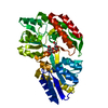
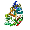
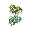
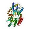
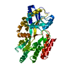
- PDB-1lls: CRYSTAL STRUCTURE OF UNLIGANDED MALTOSE BINDING PROTEIN WITH XENON -
+ Open data

- Basic information
| Entry | Database: PDB / ID: 1lls | ||||||
|---|---|---|---|---|---|---|---|
| Title | CRYSTAL STRUCTURE OF UNLIGANDED MALTOSE BINDING PROTEIN WITH XENON | ||||||
 Components Components | Maltose-binding periplasmic protein | ||||||
 Keywords Keywords | SUGAR BINDING PROTEIN / Hydrophobic Cavities / Ligand-Protein Interactions /  Xenon Binding / Xenon Derivative Xenon Binding / Xenon Derivative | ||||||
| Function / homology |  Function and homology information Function and homology informationdetection of maltose stimulus / maltose binding / maltose transport complex / maltose transport / maltodextrin transmembrane transport / carbohydrate transmembrane transporter activity / ATP-binding cassette (ABC) transporter complex, substrate-binding subunit-containing / carbohydrate transport / ATP-binding cassette (ABC) transporter complex / cell chemotaxis ...detection of maltose stimulus / maltose binding / maltose transport complex / maltose transport / maltodextrin transmembrane transport / carbohydrate transmembrane transporter activity / ATP-binding cassette (ABC) transporter complex, substrate-binding subunit-containing / carbohydrate transport / ATP-binding cassette (ABC) transporter complex / cell chemotaxis / outer membrane-bounded periplasmic space / periplasmic space / DNA damage response / membraneSimilarity search - Function | ||||||
| Biological species |  Escherichia coli (E. coli) Escherichia coli (E. coli) | ||||||
| Method | X-RAY DIFFRACTION / SYNCHROTRON / Isomorphous with Structure in Database / Resolution: 1.8 Å | ||||||
 Authors Authors | Rubin, S.M. / Lee, S.-Y. / Ruiz, E.J. / Pines, A. / Wemmer, D.E. | ||||||
 Citation Citation | Journal: J.MOL.BIOL. / Year: 2002 Title: DETECTION AND CHARACTERIZATION OF XENON-BINDING SITES IN PROTEINS BY 129XE NMR SPECTROSCOPY Authors: Rubin, S.M. / Lee, S.-Y. / Ruiz, E.J. / Pines, A. / Wemmer, D.E. #1: Journal: J.Am.Chem.Soc. / Year: 2001Title: Detection of a Conformational Change in Maltose Binding Protein by 129Xe Nuclear Magnetic Resonance Authors: Rubin, S.M. / Spence, M.M. / Dimitrov, I.E. / Ruiz, E.J. / Pines, A. / Wemmer, D.E. #2: Journal: J.Biol.Chem. / Year: 1991Title: The 2.3 A Structure of the Malto- or Maltodextrin-Binding Protein, a Primary Receptor of Bacterial Active Transport Authors: Spurlino, J.C. / Lu, G.-Y. / Quiocho, F.A. #3: Journal: Biochemistry / Year: 1992Title: Crystallographic Evidence of a Large Ligand-Induced Hinge-Twist Motion Between the Two Domains of the Maltodextrin Binding Protein Involved in Active Transport and Chemotaxis Authors: Sharff, A.J. / Rodseth, L.E. / Spurlino, J.C. / Quiocho, F.A. #4: Journal: J.Mol.Biol. / Year: 2000Title: Size Versus Polarizability in Protein-Ligand Interactions: Binding of Noble Gases within Engineered Cavities in Phage T4 Lysozyme Authors: Quillin, M.L. / Breyer, W.A. / Griswold, I.J. / Matthews, B.W. | ||||||
| History |
|
- Structure visualization
| Structure viewer | Molecule: MolmilJmol/JSmol |
|---|
- Downloads & links
-Download
| PDBx/mmCIF format | 1lls.cif.gz | 87 KB | Display | PDBx/mmCIF format |
|---|---|---|---|---|
| PDB format | pdb1lls.ent.gz | 64.6 KB | Display | PDB format |
| PDBx/mmJSON format | 1lls.json.gz | Tree view | PDBx/mmJSON format | |
| Others |  Other downloads Other downloads |
-Validation report
| Arichive directory | https://data.pdbj.org/pub/pdb/validation_reports/ll/1llsftp://data.pdbj.org/pub/pdb/validation_reports/ll/1lls | HTTPS FTP |
|---|
-Related structure data
| Related structure data |  1ompS S: Starting model for refinement |
|---|---|
| Similar structure data |


-Links
 PDBj
PDBj


- Assembly
| Deposited unit | 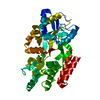
| ||||||||
|---|---|---|---|---|---|---|---|---|---|
| 1 |
| ||||||||
| Unit cell |
|
-Components
| #1: Protein | Mass: 40753.152 Da / Num. of mol.: 1 Source method: isolated from a genetically manipulated source Source: (gene. exp.) Escherichia coli (E. coli) / Plasmid: PET21a / Species (production host): Escherichia coli / Production host: Escherichia coli BL21(DE3) (bacteria) / Strain (production host): BL21(DE3) / References: UniProt: P02928, UniProt: P0AEX9*PLUS |
|---|---|
| #2: Chemical | ChemComp-XE / Xenon  Mass: 131.293 Da / Num. of mol.: 1 / Source method: obtained synthetically / Formula: Xe Mass: 131.293 Da / Num. of mol.: 1 / Source method: obtained synthetically / Formula: Xe |
| #3: Water | ChemComp-HOH / Water Mass: 18.015 Da / Num. of mol.: 171 / Source method: isolated from a natural source / Formula: H2O Mass: 18.015 Da / Num. of mol.: 171 / Source method: isolated from a natural source / Formula: H2O |
-Experimental details
-Experiment
| Experiment | Method: X-RAY DIFFRACTION / Number of used crystals: 1 |
|---|
- Sample preparation
| Crystal | Density Matthews: 2.21 Å3/Da / Density % sol: 44.31 % | ||||||||||||||||||||||||
|---|---|---|---|---|---|---|---|---|---|---|---|---|---|---|---|---|---|---|---|---|---|---|---|---|---|
| Crystal grow | Temperature: 298 K / Method: vapor diffusion, hanging drop / pH: 6.6 Details: 10 mM Sodium Citrate, 23% PEG 8000, pH 6.6, VAPOR DIFFUSION, HANGING DROP, temperature 298K | ||||||||||||||||||||||||
| Crystal grow | *PLUS | ||||||||||||||||||||||||
| Components of the solutions | *PLUS
|
-Data collection
| Diffraction | Mean temperature: 100 K |
|---|---|
| Diffraction source | Source: SYNCHROTRON / Site: ALS  / Beamline: 8.3.1 / Wavelength: 1 Å / Beamline: 8.3.1 / Wavelength: 1 Å |
| Detector | Type: ADSC / Detector: CCD / Date: Jan 31, 2002 |
| Radiation | Monochromator: Double Crystal / Protocol: SINGLE WAVELENGTH / Monochromatic (M) / Laue (L): M / Scattering type: x-ray |
| Radiation wavelength | Wavelength: 1 Å / Relative weight: 1 |
| Reflection | Resolution: 1.8→17.07 Å / Num. all: 55908 / Num. obs: 29855 / % possible obs: 92.3 % / Observed criterion σ(F): 4 / Observed criterion σ(I): 4 / Biso Wilson estimate: 16.9 Å2 / Rsym value: 0.029 / Net I/σ(I): 19.9 |
| Reflection shell | Resolution: 1.8→1.91 Å / Mean I/σ(I) obs: 10.4 / Num. unique all: 4127 / Rsym value: 0.105 / % possible all: 76 |
| Reflection | *PLUS Num. measured all: 55908 / Rmerge(I) obs: 0.029 |
- Processing
| Software |
| |||||||||||||||||||||||||
|---|---|---|---|---|---|---|---|---|---|---|---|---|---|---|---|---|---|---|---|---|---|---|---|---|---|---|
| Refinement | Method to determine structure: Isomorphous with Structure in Database Starting model: PDB ENTRY 1OMP Resolution: 1.8→17.07 Å / Rfactor Rfree error: 0.006 / Isotropic thermal model: OVERALL / Cross valid method: THROUGHOUT / σ(F): 0 / Stereochemistry target values: Engh & Huber
| |||||||||||||||||||||||||
| Solvent computation | Solvent model: FLAT MODEL / Bsol: 41.8917 Å2 / ksol: 0.387004 e/Å3 | |||||||||||||||||||||||||
| Displacement parameters | Biso mean: 22.5 Å2
| |||||||||||||||||||||||||
| Refine analyze | Luzzati coordinate error free: 0.24 Å / Luzzati sigma a free: 0.15 Å | |||||||||||||||||||||||||
| Refinement step | Cycle: LAST / Resolution: 1.8→17.07 Å
| |||||||||||||||||||||||||
| Refine LS restraints |
| |||||||||||||||||||||||||
| LS refinement shell | Resolution: 1.8→1.91 Å / Rfactor Rfree error: 0.018 / Total num. of bins used: 6
| |||||||||||||||||||||||||
| Xplor file |
| |||||||||||||||||||||||||
| Refinement | *PLUS Num. reflection obs: 29755 / Rfactor all: 0.2051 / Rfactor obs: 0.204 / Rfactor Rfree: 0.226 / Rfactor Rwork: 0.204 | |||||||||||||||||||||||||
| Solvent computation | *PLUS | |||||||||||||||||||||||||
| Displacement parameters | *PLUS | |||||||||||||||||||||||||
| Refine LS restraints | *PLUS
| |||||||||||||||||||||||||
| LS refinement shell | *PLUS Rfactor Rfree: 0.258 / Rfactor Rwork: 0.241 |
