 Movie
Movie Controller

[English] 日本語
 Yorodumi
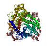
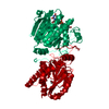
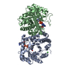
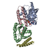
- PDB-1lf9: CRYSTAL STRUCTURE OF BACTERIAL GLUCOAMYLASE COMPLEXED WITH ACARBOSE -
+ Open data

- Basic information
| Entry | Database: PDB / ID: 1lf9 | |||||||||
|---|---|---|---|---|---|---|---|---|---|---|
| Title | CRYSTAL STRUCTURE OF BACTERIAL GLUCOAMYLASE COMPLEXED WITH ACARBOSE | |||||||||
 Components Components | GLUCOAMYLASE Glucan 1,4-a-glucosidase Glucan 1,4-a-glucosidase | |||||||||
 Keywords Keywords | HYDROLASE / (alpha/alpha) barrel / 6 alpha-helical hairpin torroid / super beta sandwich / carbohydrase family GH15 / acarbose | |||||||||
| Function / homology |  Function and homology information Function and homology informationglucan 1,4-alpha-glucosidase / glucan 1,4-alpha-glucosidase activity / carbohydrate binding / carbohydrate metabolic processSimilarity search - Function | |||||||||
| Biological species |  Thermoanaerobacterium thermosaccharolyticum (bacteria) Thermoanaerobacterium thermosaccharolyticum (bacteria) | |||||||||
| Method | X-RAY DIFFRACTION / SYNCHROTRON / MOLECULAR REPLACEMENT / Resolution: 2.2 Å | |||||||||
 Authors Authors | Aleshin, A.E. / Feng, P.-H. / Honzatko, R.B. / Reilly, P.J. | |||||||||
 Citation Citation | Journal: J.Mol.Biol. / Year: 2003 Title: Crystal structure and evolution of prokaryotic glucoamylase Authors: Aleshin, A.E. / Feng, P.-H. / Honzatko, R.B. / Reilly, P.J. | |||||||||
| History |
| |||||||||
| Remark 999 | SEQUENCE Author states the sequence of this crystal structure differs from the DNA sequence of ...SEQUENCE Author states the sequence of this crystal structure differs from the DNA sequence of GenBank entry AAC24003. The crystal structure has insertions at residue 125 and after residue 679. These differences are consistent with Genbank entry BAA02251. |
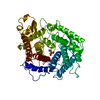
- Structure visualization
| Structure viewer | Molecule: MolmilJmol/JSmol |
|---|
- Downloads & links
-Download
| PDBx/mmCIF format | 1lf9.cif.gz | 288.4 KB | Display | PDBx/mmCIF format |
|---|---|---|---|---|
| PDB format | pdb1lf9.ent.gz | 230.3 KB | Display | PDB format |
| PDBx/mmJSON format | 1lf9.json.gz | Tree view | PDBx/mmJSON format | |
| Others |  Other downloads Other downloads |
-Validation report
| Arichive directory | https://data.pdbj.org/pub/pdb/validation_reports/lf/1lf9ftp://data.pdbj.org/pub/pdb/validation_reports/lf/1lf9 | HTTPS FTP |
|---|
-Related structure data
| Related structure data | 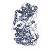 1lf6SC S: Starting model for refinement C: citing same article ( |
|---|---|
| Similar structure data |


-Links
 PDBj
PDBj


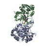
- Assembly
| Deposited unit | 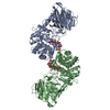
| |||||||||
|---|---|---|---|---|---|---|---|---|---|---|
| 1 | 
| |||||||||
| 2 | 
| |||||||||
| Unit cell |
| |||||||||
| Noncrystallographic symmetry (NCS) | NCS domain:
| |||||||||
| Details | The presumable biological assembly is a monomer, but the crystallographically observed dimer may also be functional |
-Components
| #1: Protein | Glucan 1,4-a-glucosidase / GLUCAN 1 / 4-ALPHA-GLUCOSIDASE / 1 / 4-ALPHA-D-GLUCAN GLUCOHYDROLASE / AMYLOGLUCOSIDASE / GAMMA- ...GLUCAN 1 / 4-ALPHA-GLUCOSIDASE / 1 / 4-ALPHA-D-GLUCAN GLUCOHYDROLASE / AMYLOGLUCOSIDASE / GAMMA-AMYLASE / LYSOSOMAL ALPHA-GLUCOSIDASE / EXO-1 / 4-ALPHA-GLUCOSIDASE Mass: 76590.500 Da / Num. of mol.: 2 / Source method: isolated from a natural source Source: (natural) Thermoanaerobacterium thermosaccharolyticum (bacteria)Strain: DSM 571 References: GenBank: 3243238, UniProt: O85672*PLUS, glucan 1,4-alpha-glucosidase #2: Polysaccharide | 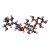  Type: oligosaccharide, Oligosaccharide / Class: Inhibitor / Mass: 645.606 Da / Num. of mol.: 2 Type: oligosaccharide, Oligosaccharide / Class: Inhibitor / Mass: 645.606 Da / Num. of mol.: 2Source method: isolated from a genetically manipulated source Details: oligosaccharide / References: alpha-acarbose #3: Chemical | Sulfate  Mass: 96.063 Da / Num. of mol.: 2 / Source method: obtained synthetically / Formula: SO4 Mass: 96.063 Da / Num. of mol.: 2 / Source method: obtained synthetically / Formula: SO4#4: Water | ChemComp-HOH / | Water Mass: 18.015 Da / Num. of mol.: 674 / Source method: isolated from a natural source / Formula: H2O Mass: 18.015 Da / Num. of mol.: 674 / Source method: isolated from a natural source / Formula: H2O |
|---|
-Experimental details
-Experiment
| Experiment | Method: X-RAY DIFFRACTION / Number of used crystals: 1 |
|---|
- Sample preparation
| Crystal | Density Matthews: 2.23 Å3/Da / Density % sol: 44.75 % | |||||||||||||||||||||||||||||||||||
|---|---|---|---|---|---|---|---|---|---|---|---|---|---|---|---|---|---|---|---|---|---|---|---|---|---|---|---|---|---|---|---|---|---|---|---|---|
| Crystal grow | Temperature: 298 K / Method: vapor diffusion, hanging drop / pH: 7.5 Details: PEG 3350, Tris-HCl, Lithium Sulfate, pH 7.5, VAPOR DIFFUSION, HANGING DROP, temperature 298K | |||||||||||||||||||||||||||||||||||
| Crystal grow | *PLUS Temperature: 24 ℃ | |||||||||||||||||||||||||||||||||||
| Components of the solutions | *PLUS
|
-Data collection
| Diffraction | Mean temperature: 100 K |
|---|---|
| Diffraction source | Source: SYNCHROTRON / Site: APS  / Beamline: 19-BM / Wavelength: 1 Å / Beamline: 19-BM / Wavelength: 1 Å |
| Detector | Type: ADSC QUANTUM 4 / Detector: CCD / Date: Apr 5, 2001 |
| Radiation | Monochromator: Si 111 / Protocol: SINGLE WAVELENGTH / Monochromatic (M) / Laue (L): M / Scattering type: x-ray |
| Radiation wavelength | Wavelength: 1 Å / Relative weight: 1 |
| Reflection | Resolution: 2.2→30 Å / Num. all: 70153 / Num. obs: 65095 / % possible obs: 92 % / Observed criterion σ(F): 0 / Observed criterion σ(I): 0 / Redundancy: 4 % / Biso Wilson estimate: 35.5 Å2 / Rmerge(I) obs: 0.11 / Net I/σ(I): 14 |
| Reflection shell | Resolution: 2.2→2.3 Å / Rmerge(I) obs: 0.4 / % possible all: 83 |
| Reflection | *PLUS Highest resolution: 2.2 Å / Lowest resolution: 30 Å / % possible obs: 92 % / Num. measured all: 268721 |
| Reflection shell | *PLUS % possible obs: 83 % / Rmerge(I) obs: 0.4 |
- Processing
| Software |
| ||||||||||||||||||||||||||||||||||||
|---|---|---|---|---|---|---|---|---|---|---|---|---|---|---|---|---|---|---|---|---|---|---|---|---|---|---|---|---|---|---|---|---|---|---|---|---|---|
| Refinement | Method to determine structure: MOLECULAR REPLACEMENT Starting model: PDB ENTRY 1LF6 Resolution: 2.2→29.86 Å / Rfactor Rfree error: 0.003 / Data cutoff high absF: 2983770.37 / Data cutoff high rms absF: 2983770.37 / Data cutoff low absF: 0 / Isotropic thermal model: RESTRAINED / Cross valid method: THROUGHOUT / σ(F): 0 / Stereochemistry target values: Engh & Huber
| ||||||||||||||||||||||||||||||||||||
| Solvent computation | Solvent model: FLAT MODEL / Bsol: 49.0398 Å2 / ksol: 0.348347 e/Å3 | ||||||||||||||||||||||||||||||||||||
| Displacement parameters | Biso mean: 36.2 Å2
| ||||||||||||||||||||||||||||||||||||
| Refine analyze |
| ||||||||||||||||||||||||||||||||||||
| Refinement step | Cycle: LAST / Resolution: 2.2→29.86 Å
| ||||||||||||||||||||||||||||||||||||
| Refine LS restraints |
| ||||||||||||||||||||||||||||||||||||
| Refine LS restraints NCS |
| ||||||||||||||||||||||||||||||||||||
| LS refinement shell | Resolution: 2.2→2.34 Å / Rfactor Rfree error: 0.011 / Total num. of bins used: 6
| ||||||||||||||||||||||||||||||||||||
| Xplor file |
| ||||||||||||||||||||||||||||||||||||
| Refinement | *PLUS Highest resolution: 2.2 Å / Lowest resolution: 30 Å / % reflection Rfree: 7 % / Rfactor Rfree: 0.23 / Rfactor Rwork: 0.19 | ||||||||||||||||||||||||||||||||||||
| Solvent computation | *PLUS | ||||||||||||||||||||||||||||||||||||
| Displacement parameters | *PLUS | ||||||||||||||||||||||||||||||||||||
| Refine LS restraints | *PLUS
|
