 Movie
Movie Controller

[English] 日本語
 Yorodumi
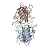
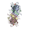
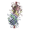
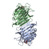
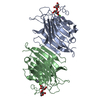
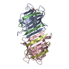
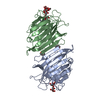
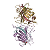
- PDB-1lem: THE MONOSACCHARIDE BINDING SITE OF LENTIL LECTIN: AN X-RAY AND MO... -
+ Open data

- Basic information
| Entry | Database: PDB / ID: 1lem | ||||||
|---|---|---|---|---|---|---|---|
| Title | THE MONOSACCHARIDE BINDING SITE OF LENTIL LECTIN: AN X-RAY AND MOLECULAR MODELLING STUDY | ||||||
 Components Components | (LECTIN ) x 2 ) x 2 | ||||||
 Keywords Keywords | LECTIN | ||||||
| Function / homology |  Function and homology information Function and homology informationcarbohydrate mediated signaling / D-mannose binding / manganese ion binding / carbohydrate binding / calcium ion bindingSimilarity search - Function | ||||||
| Biological species |  Lens culinaris (lentil) Lens culinaris (lentil) | ||||||
| Method | X-RAY DIFFRACTION / Resolution: 3 Å | ||||||
 Authors Authors | Loris, R. / Wyns, L. | ||||||
 Citation Citation | Journal: Glycoconj.J. / Year: 1994 Title: The monosaccharide binding site of lentil lectin: an X-ray and molecular modelling study. Authors: Loris, R. / Casset, F. / Bouckaert, J. / Pletinckx, J. / Dao-Thi, M.H. / Poortmans, F. / Imberty, A. / Perez, S. / Wyns, L. #1: Journal: Biochemistry / Year: 1993Title: Crystal Structure Determination and Refinement at 2.3 Angstroms Resolution of the Lentil Lectin Authors: Loris, R. / Steyaert, J. / Maes, D. / Lisgarten, J. / Pickersgill, R. / Wyns, L. #2: Journal: J.Mol.Biol. / Year: 1992Title: Two Crystal Forms of the Lentil Lectin Diffract to High Resolution Authors: Loris, R. / Lisgarten, J. / Maes, D. / Pickersgill, R. / Korber, F. / Reynolds, C. / Wyns, L. | ||||||
| History |
|
- Structure visualization
| Structure viewer | Molecule: MolmilJmol/JSmol |
|---|
- Downloads & links
-Download
| PDBx/mmCIF format | 1lem.cif.gz | 58.3 KB | Display | PDBx/mmCIF format |
|---|---|---|---|---|
| PDB format | pdb1lem.ent.gz | 42.4 KB | Display | PDB format |
| PDBx/mmJSON format | 1lem.json.gz | Tree view | PDBx/mmJSON format | |
| Others |  Other downloads Other downloads |
-Validation report
| Arichive directory | https://data.pdbj.org/pub/pdb/validation_reports/le/1lemftp://data.pdbj.org/pub/pdb/validation_reports/le/1lem | HTTPS FTP |
|---|
-Related structure data
| Similar structure data |
|---|


-Links
 PDBj
PDBj


- Assembly
| Deposited unit | 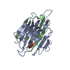
| ||||||||
|---|---|---|---|---|---|---|---|---|---|
| 1 | 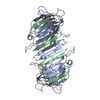
| ||||||||
| Unit cell |
| ||||||||
| Atom site foot note | 1: ALA A 80 - ASP A 81 OMEGA = 0.73 PEPTIDE BOND DEVIATES SIGNIFICANTLY FROM TRANS CONFORMATION |
-Components
-Protein , 2 types, 2 molecules AB
| #1: Protein | Mass: 19906.982 Da / Num. of mol.: 1 Source method: isolated from a genetically manipulated source Source: (gene. exp.) Lens culinaris (lentil) / References: UniProt: P02870 |
|---|---|
| #2: Protein | Mass: 5714.288 Da / Num. of mol.: 1 Source method: isolated from a genetically manipulated source Source: (gene. exp.) Lens culinaris (lentil) / References: UniProt: P02870 |
-Sugars , 1 types, 1 molecules 
| #3: Sugar | ChemComp-GLC / Glucose Type: D-saccharide, alpha linking / Mass: 180.156 Da / Num. of mol.: 1 Type: D-saccharide, alpha linking / Mass: 180.156 Da / Num. of mol.: 1Source method: isolated from a genetically manipulated source Formula: C6H12O6 |
|---|
-Non-polymers , 3 types, 45 molecules 


| #4: Chemical | ChemComp-CA /  Mass: 40.078 Da / Num. of mol.: 1 / Source method: obtained synthetically / Formula: Ca Mass: 40.078 Da / Num. of mol.: 1 / Source method: obtained synthetically / Formula: Ca |
|---|---|
| #5: Chemical | ChemComp-MN /  Mass: 54.938 Da / Num. of mol.: 1 / Source method: obtained synthetically / Formula: Mn Mass: 54.938 Da / Num. of mol.: 1 / Source method: obtained synthetically / Formula: Mn |
| #6: Water | ChemComp-HOH / WaterMass: 18.015 Da / Num. of mol.: 43 / Source method: isolated from a natural source / Formula: H2O |
-Details
| Nonpolymer details | THE LENTIL LECTIN MONOMER HAS A BOUND CALCIUM AND MANGANESE ION. THE CALCIUM AND MANGANESE IONS ARE ...THE LENTIL LECTIN MONOMER HAS A BOUND CALCIUM AND MANGANESE ION. THE CALCIUM AND MANGANESE IONS ARE ESSENTIAL FOR STABILIZIN |
|---|
-Experimental details
-Experiment
| Experiment | Method: X-RAY DIFFRACTION |
|---|
- Sample preparation
| Crystal | Density Matthews: 3.42 Å3/Da / Density % sol: 64.04 % | |||||||||||||||||||||||||||||||||||
|---|---|---|---|---|---|---|---|---|---|---|---|---|---|---|---|---|---|---|---|---|---|---|---|---|---|---|---|---|---|---|---|---|---|---|---|---|
| Crystal grow | *PLUS pH: 6.5 / Method: vapor diffusion, hanging drop | |||||||||||||||||||||||||||||||||||
| Components of the solutions | *PLUS
|
-Data collection
| Radiation | Scattering type: x-ray |
|---|---|
| Radiation wavelength | Relative weight: 1 |
| Reflection | *PLUS Highest resolution: 3 Å / Lowest resolution: 10 Å / Num. obs: 6950 / % possible obs: 94.5 % / Num. measured all: 35751 / Rmerge(I) obs: 0.07 |
- Processing
| Software |
| ||||||||||||||||||||||||||||||||||||||||||||||||||||||||||||
|---|---|---|---|---|---|---|---|---|---|---|---|---|---|---|---|---|---|---|---|---|---|---|---|---|---|---|---|---|---|---|---|---|---|---|---|---|---|---|---|---|---|---|---|---|---|---|---|---|---|---|---|---|---|---|---|---|---|---|---|---|---|
| Refinement | Rfactor Rwork: 0.206 / Rfactor obs: 0.206 / Highest resolution: 3 Å | ||||||||||||||||||||||||||||||||||||||||||||||||||||||||||||
| Refinement step | Cycle: LAST / Highest resolution: 3 Å
| ||||||||||||||||||||||||||||||||||||||||||||||||||||||||||||
| Refine LS restraints |
| ||||||||||||||||||||||||||||||||||||||||||||||||||||||||||||
| Software | *PLUS Name: 'X-PLOR' / Classification: refinement | ||||||||||||||||||||||||||||||||||||||||||||||||||||||||||||
| Refinement | *PLUS Lowest resolution: 10 Å | ||||||||||||||||||||||||||||||||||||||||||||||||||||||||||||
| Solvent computation | *PLUS | ||||||||||||||||||||||||||||||||||||||||||||||||||||||||||||
| Displacement parameters | *PLUS | ||||||||||||||||||||||||||||||||||||||||||||||||||||||||||||
| Refine LS restraints | *PLUS
|
