 Movie
Movie Controller

[English] 日本語
 Yorodumi
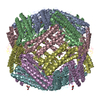
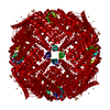
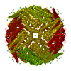
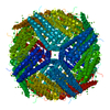
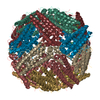
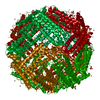
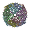
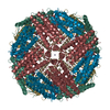
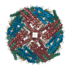
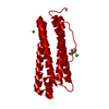
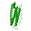
- PDB-1lb3: Structure of recombinant mouse L chain ferritin at 1.2 A resolution -
+ Open data

- Basic information
| Entry | Database: PDB / ID: 1lb3 | ||||||
|---|---|---|---|---|---|---|---|
| Title | Structure of recombinant mouse L chain ferritin at 1.2 A resolution | ||||||
 Components Components | FERRITIN LIGHT CHAIN 1 | ||||||
 Keywords Keywords | METAL BINDING PROTEIN / IRON STORAGE | ||||||
| Function / homology |  Function and homology information Function and homology information: / autolysosome / intracellular sequestering of iron ion / endocytic vesicle lumen / ferric iron binding / ferrous iron binding / iron ion transport / iron ion binding / extracellular region / identical protein binding / cytoplasmSimilarity search - Function | ||||||
| Biological species |  Mus musculus (house mouse) Mus musculus (house mouse) | ||||||
| Method | X-RAY DIFFRACTION / SYNCHROTRON / MOLECULAR REPLACEMENT / Resolution: 1.21 Å | ||||||
 Authors Authors | Granier, T. / Langlois D'Estaintot, B. / Gallois, B. / Chevalier, J.-M. / Precigoux, G. / Santambrogio, P. / Arosio, P. | ||||||
 Citation Citation | Journal: J.Biol.Inorg.Chem. / Year: 2003 Title: Structural description of the active sites of mouse L-chain ferritin at 1.2A resolution Authors: Granier, T. / Langlois D'Estaintot, B. / Gallois, B. / Chevalier, J.-M. / Precigoux, G. / Santambrogio, P. / Arosio, P. | ||||||
| History |
|
- Structure visualization
| Structure viewer | Molecule: MolmilJmol/JSmol |
|---|
- Downloads & links
-Download
| PDBx/mmCIF format | 1lb3.cif.gz | 106.4 KB | Display | PDBx/mmCIF format |
|---|---|---|---|---|
| PDB format | pdb1lb3.ent.gz | 80.4 KB | Display | PDB format |
| PDBx/mmJSON format | 1lb3.json.gz | Tree view | PDBx/mmJSON format | |
| Others |  Other downloads Other downloads |
-Validation report
| Arichive directory | https://data.pdbj.org/pub/pdb/validation_reports/lb/1lb3ftp://data.pdbj.org/pub/pdb/validation_reports/lb/1lb3 | HTTPS FTP |
|---|
-Related structure data
| Related structure data |  1h96S S: Starting model for refinement |
|---|---|
| Similar structure data |


-Links
 PDBj
PDBj


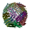
- Assembly
| Deposited unit | 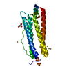
| ||||||||
|---|---|---|---|---|---|---|---|---|---|
| 1 | x 24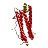
| ||||||||
| Unit cell |
| ||||||||
| Components on special symmetry positions |
| ||||||||
| Details | The biological assembly is a 24mer. Coordinates for a complete multimer representing the known biologically significant oligomerization state of the molecule can be generated from the monomer in the assymmetric unit by the following symmetry operations: -X,-Y,Z; -X,Y,-Z; X,-Y,-Z; Z,X,Y; Z,-X,-Y; -Z,-X,Y; -Z,X,-Y; Y,Z,X; -Y,Z,-X; Y,-Z,-X; -Y,-Z,X; Y,X,-Z; -Y,-X,-Z; Y,-X,Z; -Y,X,Z; X,Z,-Y; -X,Z,Y; -X,-Z,-Y; X,-Z,Y; Z,Y,-X; Z,-Y,X; -Z,Y,X; -Z,-Y,-X; |
-Components
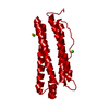
| #1: Protein | / FERRITIN L SUBUNIT 1 Mass: 20670.164 Da / Num. of mol.: 1 / Mutation: T121A Source method: isolated from a genetically manipulated source Source: (gene. exp.) Mus musculus (house mouse) / Gene: FTL / Plasmid: pMLF27 / Species (production host): Escherichia coli / Production host:  Escherichia coli BL21(DE3) (bacteria) / Strain (production host): BL21-DE3 / References: UniProt: P29391 Escherichia coli BL21(DE3) (bacteria) / Strain (production host): BL21-DE3 / References: UniProt: P29391 | ||||||||
|---|---|---|---|---|---|---|---|---|---|
| #2: Chemical | Sulfate  Mass: 96.063 Da / Num. of mol.: 2 / Source method: obtained synthetically / Formula: SO4 Mass: 96.063 Da / Num. of mol.: 2 / Source method: obtained synthetically / Formula: SO4#3: Chemical | ChemComp-CD /   Mass: 112.411 Da / Num. of mol.: 9 / Source method: obtained synthetically / Formula: Cd Mass: 112.411 Da / Num. of mol.: 9 / Source method: obtained synthetically / Formula: Cd#4: Chemical | Glycerol  Mass: 92.094 Da / Num. of mol.: 2 / Source method: obtained synthetically / Formula: C3H8O3 Mass: 92.094 Da / Num. of mol.: 2 / Source method: obtained synthetically / Formula: C3H8O3#5: Water | ChemComp-HOH / | Water Mass: 18.015 Da / Num. of mol.: 286 / Source method: isolated from a natural source / Formula: H2O Mass: 18.015 Da / Num. of mol.: 286 / Source method: isolated from a natural source / Formula: H2OCompound details | CHAIN ENGINEERED | |
-Experimental details
-Experiment
| Experiment | Method: X-RAY DIFFRACTION / Number of used crystals: 1 |
|---|
- Sample preparation
| Crystal | Density Matthews: 2.72 Å3/Da / Density % sol: 54.8 % | ||||||||||||||||||||||||||||||||||||||||||
|---|---|---|---|---|---|---|---|---|---|---|---|---|---|---|---|---|---|---|---|---|---|---|---|---|---|---|---|---|---|---|---|---|---|---|---|---|---|---|---|---|---|---|---|
| Crystal grow | Temperature: 290 K / Method: vapor diffusion, hanging drop / pH: 7.4 Details: ammonium sulphate, cadmium sulphate, sodium azide, tris HCl, pH 7.4, VAPOR DIFFUSION, HANGING DROP, temperature 290K | ||||||||||||||||||||||||||||||||||||||||||
| Crystal grow | *PLUS Temperature: 293 K / Details: Granier, T., (2001) Acta Crystallogr., D57, 1491. | ||||||||||||||||||||||||||||||||||||||||||
| Components of the solutions | *PLUS
|
-Data collection
| Diffraction | Mean temperature: 150 K |
|---|---|
| Diffraction source | Source: SYNCHROTRON / Site: LURE  / Beamline: DW32 / Wavelength: 0.966 / Wavelength: 0.966 Å / Beamline: DW32 / Wavelength: 0.966 / Wavelength: 0.966 Å |
| Detector | Type: MARRESEARCH / Detector: IMAGE PLATE / Date: Apr 17, 2000 / Details: W/SI MIRRORS |
| Radiation | Monochromator: Si (111) CHANNEL / Protocol: SINGLE WAVELENGTH / Monochromatic (M) / Laue (L): M / Scattering type: x-ray |
| Radiation wavelength | Wavelength: 0.966 Å / Relative weight: 1 |
| Reflection | Resolution: 1.21→14 Å / Num. all: 74509 / Num. obs: 74509 / % possible obs: 98.4 % / Observed criterion σ(F): 0 / Observed criterion σ(I): 0 / Redundancy: 5.5 % / Rsym value: 0.066 / Net I/σ(I): 6.4 |
| Reflection shell | Resolution: 1.21→1.25 Å / Redundancy: 4.7 % / Mean I/σ(I) obs: 2 / Num. unique all: 5144 / Rsym value: 0.48 / % possible all: 98.4 |
| Reflection | *PLUS Lowest resolution: 14 Å / Num. measured all: 399787 / Rmerge(I) obs: 0.078 |
| Reflection shell | *PLUS % possible obs: 98.4 % / Num. unique obs: 5144 / Rmerge(I) obs: 0.602 |
- Processing
| Software |
| |||||||||||||||||||||||||||||||||
|---|---|---|---|---|---|---|---|---|---|---|---|---|---|---|---|---|---|---|---|---|---|---|---|---|---|---|---|---|---|---|---|---|---|---|
| Refinement | Method to determine structure: MOLECULAR REPLACEMENT Starting model: PDB ENTRY 1H96 Resolution: 1.21→14 Å / Num. parameters: 16266 / Num. restraintsaints: 16802 / Isotropic thermal model: ANISOTROPIC / Cross valid method: FREE R / σ(F): 0 / σ(I): 0 / Stereochemistry target values: Engh & Huber / Details: ANISOTROPIC REFINEMENT REDUCED FREE R (NO CUTOFF)
| |||||||||||||||||||||||||||||||||
| Solvent computation | Solvent model: MOEWS & KRETSINGER, J.MOL.BIOL.91(1973)201-22 | |||||||||||||||||||||||||||||||||
| Refine analyze | Num. disordered residues: 38 / Occupancy sum hydrogen: 1287.25 / Occupancy sum non hydrogen: 1622.33 | |||||||||||||||||||||||||||||||||
| Refinement step | Cycle: LAST / Resolution: 1.21→14 Å
| |||||||||||||||||||||||||||||||||
| Refine LS restraints |
| |||||||||||||||||||||||||||||||||
| LS refinement shell | Resolution: 1.21→1.26 Å
| |||||||||||||||||||||||||||||||||
| Software | *PLUS Name: SHELXL / Version: 97 / Classification: refinement | |||||||||||||||||||||||||||||||||
| Refinement | *PLUS Lowest resolution: 14 Å / Rfactor all: 0.134 / Rfactor Rfree: 0.16 | |||||||||||||||||||||||||||||||||
| Solvent computation | *PLUS | |||||||||||||||||||||||||||||||||
| Displacement parameters | *PLUS | |||||||||||||||||||||||||||||||||
| Refine LS restraints | *PLUS
| |||||||||||||||||||||||||||||||||
| LS refinement shell | *PLUS Rfactor Rfree: 0.22 / Rfactor Rwork: 0.2 |
