 Movie
Movie Controller

[English] 日本語
 Yorodumi
- PDB-1l9e: Role of Histidine 269 in Catalysis by Monomeric Sarcosine Oxidase -
+ Open data

- Basic information
| Entry | Database: PDB / ID: 1l9e | ||||||
|---|---|---|---|---|---|---|---|
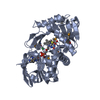
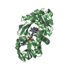
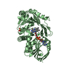
| Title | Role of Histidine 269 in Catalysis by Monomeric Sarcosine Oxidase | ||||||
 Components Components | Monomeric sarcosine oxidase | ||||||
 Keywords Keywords |  OXIDOREDUCTASE / flavoprotein / oxidase OXIDOREDUCTASE / flavoprotein / oxidase | ||||||
| Function / homology |  Function and homology information Function and homology informationL-lysine catabolic process to acetyl-CoA via L-pipecolate / L-pipecolate oxidase activity / sarcosine oxidase (formaldehyde-forming) / saccharopine oxidase activity / sarcosine oxidase activity / peroxisome / flavin adenine dinucleotide binding / cytosolSimilarity search - Function | ||||||
| Biological species |  | ||||||
| Method | X-RAY DIFFRACTION / refined directly / Resolution: 1.85 Å | ||||||
 Authors Authors | Zhao, G. / Song, H. / Chen, Z.-w. / Mathews, F.S. / Jorns, M.S. | ||||||
 Citation Citation | Journal: Biochemistry / Year: 2002 Title: Monomeric sarcosine oxidase: role of histidine 269 in catalysis. Authors: Zhao, G. / Song, H. / Chen, Z.W. / Mathews, F.S. / Jorns, M.S. #1: Journal: Structure / Year: 1999Title: Monomeric sarcosine oxidase: structure of a covalently flavinylated amine oxidizing enzyme Authors: Trickey, P. / Wagner, M.A. / Jorns, M.S. / Mathews, F.S. | ||||||
| History |
|
- Structure visualization
| Structure viewer | Molecule: MolmilJmol/JSmol |
|---|
- Downloads & links
-Download
| PDBx/mmCIF format | 1l9e.cif.gz | 176.7 KB | Display | PDBx/mmCIF format |
|---|---|---|---|---|
| PDB format | pdb1l9e.ent.gz | 137.6 KB | Display | PDB format |
| PDBx/mmJSON format | 1l9e.json.gz | Tree view | PDBx/mmJSON format | |
| Others |  Other downloads Other downloads |
-Validation report
| Arichive directory | https://data.pdbj.org/pub/pdb/validation_reports/l9/1l9eftp://data.pdbj.org/pub/pdb/validation_reports/l9/1l9e | HTTPS FTP |
|---|
-Related structure data
| Related structure data |  1l9cC  1l9dC  1b3m C: citing same article ( S: Starting model for refinement |
|---|---|
| Similar structure data |


-Links
 PDBj
PDBj


- Assembly
| Deposited unit | 
| ||||||||||
|---|---|---|---|---|---|---|---|---|---|---|---|
| 1 | 
| ||||||||||
| 2 | 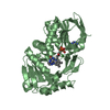
| ||||||||||
| Unit cell |
| ||||||||||
| Details | The biological assembly is monomer. |
-Components
| #1: Protein | Mass: 43101.355 Da / Num. of mol.: 2 Source method: isolated from a genetically manipulated source Source: (gene. exp.) Escherichia coli (E. coli) / Strain (production host): DH1References: UniProt: P40859, sarcosine oxidase (formaldehyde-forming) #2: Chemical | Chloride  Mass: 35.453 Da / Num. of mol.: 2 / Source method: obtained synthetically / Formula: Cl Mass: 35.453 Da / Num. of mol.: 2 / Source method: obtained synthetically / Formula: Cl#3: Chemical | Flavin adenine dinucleotide  Mass: 785.550 Da / Num. of mol.: 2 / Source method: obtained synthetically / Formula: C27H33N9O15P2 / Comment: FAD*YM Mass: 785.550 Da / Num. of mol.: 2 / Source method: obtained synthetically / Formula: C27H33N9O15P2 / Comment: FAD*YM#4: Chemical | Imidazole  Mass: 69.085 Da / Num. of mol.: 2 / Source method: obtained synthetically / Formula: C3H5N2 Mass: 69.085 Da / Num. of mol.: 2 / Source method: obtained synthetically / Formula: C3H5N2#5: Water | ChemComp-HOH / | Water Mass: 18.015 Da / Num. of mol.: 457 / Source method: isolated from a natural source / Formula: H2O Mass: 18.015 Da / Num. of mol.: 457 / Source method: isolated from a natural source / Formula: H2O |
|---|
-Experimental details
-Experiment
| Experiment | Method: X-RAY DIFFRACTION / Number of used crystals: 1 |
|---|
- Sample preparation
| Crystal | Density Matthews: 2.22 Å3/Da / Density % sol: 44.57 % | ||||||||||||||||||||||||
|---|---|---|---|---|---|---|---|---|---|---|---|---|---|---|---|---|---|---|---|---|---|---|---|---|---|
| Crystal grow | Temperature: 295 K / Method: vapor diffusion, sitting drop / pH: 7 Details: phosphate, imidazole, pH 7.0, VAPOR DIFFUSION, SITTING DROP at 295K, temperature 295.0K | ||||||||||||||||||||||||
| Crystal grow | *PLUS pH: 8 | ||||||||||||||||||||||||
| Components of the solutions | *PLUS
|
-Data collection
| Diffraction | Mean temperature: 298 K |
|---|---|
| Diffraction source | Source: ROTATING ANODE / Type: RIGAKU RU200 / Wavelength: 1.5418 Å |
| Detector | Type: RIGAKU RAXIS IV / Detector: IMAGE PLATE / Date: Mar 12, 2001 |
| Radiation | Monochromator: mirror / Protocol: SINGLE WAVELENGTH / Monochromatic (M) / Laue (L): M / Scattering type: x-ray |
| Radiation wavelength | Wavelength: 1.5418 Å / Relative weight: 1 |
| Reflection | Resolution: 1.85→30 Å / Num. all: 64360 / Num. obs: 57602 / % possible obs: 89.5 % / Observed criterion σ(F): 0 / Observed criterion σ(I): 0 / Redundancy: 2.9 % / Biso Wilson estimate: 13.9 Å2 / Rmerge(I) obs: 0.072 / Net I/σ(I): 14.2 |
| Reflection shell | Resolution: 1.85→1.92 Å / Redundancy: 1.7 % / Rmerge(I) obs: 0.28 / Mean I/σ(I) obs: 3 / Num. unique all: 3709 / % possible all: 57.7 |
| Reflection | *PLUS Lowest resolution: 30 Å / Rmerge(I) obs: 0.072 |
| Reflection shell | *PLUS % possible obs: 57.7 % / Rmerge(I) obs: 0.28 |
- Processing
| Software |
| |||||||||||||||||||||||||||||||||||||||||||||||||
|---|---|---|---|---|---|---|---|---|---|---|---|---|---|---|---|---|---|---|---|---|---|---|---|---|---|---|---|---|---|---|---|---|---|---|---|---|---|---|---|---|---|---|---|---|---|---|---|---|---|---|
| Refinement | Method to determine structure: refined directly Starting model: PDB code 1B3M 1b3m Resolution: 1.85→30 Å / Isotropic thermal model: Isotropic / Cross valid method: THROUGHOUT / σ(F): 0 / σ(I): 0 / Stereochemistry target values: Engh & Huber
| |||||||||||||||||||||||||||||||||||||||||||||||||
| Displacement parameters | Biso mean: 23.9 Å2 | |||||||||||||||||||||||||||||||||||||||||||||||||
| Refine analyze |
| |||||||||||||||||||||||||||||||||||||||||||||||||
| Refinement step | Cycle: LAST / Resolution: 1.85→30 Å
| |||||||||||||||||||||||||||||||||||||||||||||||||
| Refine LS restraints |
| |||||||||||||||||||||||||||||||||||||||||||||||||
| LS refinement shell |
| |||||||||||||||||||||||||||||||||||||||||||||||||
| Refinement | *PLUS Lowest resolution: 30 Å / % reflection Rfree: 10 % / Rfactor Rfree: 0.195 / Rfactor Rwork: 0.168 | |||||||||||||||||||||||||||||||||||||||||||||||||
| Solvent computation | *PLUS | |||||||||||||||||||||||||||||||||||||||||||||||||
| Displacement parameters | *PLUS | |||||||||||||||||||||||||||||||||||||||||||||||||
| Refine LS restraints | *PLUS
| |||||||||||||||||||||||||||||||||||||||||||||||||
| LS refinement shell | *PLUS Lowest resolution: 1.9 Å / Rfactor Rfree: 0.262 / Rfactor Rwork: 0.251 |
