 Movie
Movie Controller

[English] 日本語
 Yorodumi
- PDB-1l8l: Molecular basis for the local confomational rearrangement of huma... -
+ Open data

- Basic information
| Entry | Database: PDB / ID: 1l8l | ||||||
|---|---|---|---|---|---|---|---|
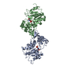
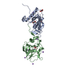
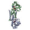
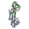
| Title | Molecular basis for the local confomational rearrangement of human phosphoserine phosphatase | ||||||
 Components Components | L-3-phosphoserine phosphatase | ||||||
 Keywords Keywords |  HYDROLASE / phosphatase / conformational rearrangement HYDROLASE / phosphatase / conformational rearrangement | ||||||
| Function / homology |  Function and homology informationphosphoserine phosphatase / Serine biosynthesis / L-phosphoserine phosphatase activity / L-serine metabolic process / L-serine biosynthetic process / response to testosterone / response to mechanical stimulus / dephosphorylation / response to nutrient levels / in utero embryonic development ...phosphoserine phosphatase / Serine biosynthesis / L-phosphoserine phosphatase activity / L-serine metabolic process / L-serine biosynthetic process / response to testosterone / response to mechanical stimulus / dephosphorylation / response to nutrient levels / in utero embryonic development / magnesium ion binding / protein homodimerization activity / identical protein binding / cytosol / cytoplasm Function and homology informationphosphoserine phosphatase / Serine biosynthesis / L-phosphoserine phosphatase activity / L-serine metabolic process / L-serine biosynthetic process / response to testosterone / response to mechanical stimulus / dephosphorylation / response to nutrient levels / in utero embryonic development ...phosphoserine phosphatase / Serine biosynthesis / L-phosphoserine phosphatase activity / L-serine metabolic process / L-serine biosynthetic process / response to testosterone / response to mechanical stimulus / dephosphorylation / response to nutrient levels / in utero embryonic development / magnesium ion binding / protein homodimerization activity / identical protein binding / cytosol / cytoplasmSimilarity search - Function | ||||||
| Biological species |  Homo sapiens (human) Homo sapiens (human) | ||||||
| Method | X-RAY DIFFRACTION / SYNCHROTRON / MAD / Resolution: 2.51 Å | ||||||
 Authors Authors | Kim, H.Y. / Heo, Y.S. / Kim, J.H. / Park, M.H. / Moon, J. / Park, S.Y. / Lee, T.G. / Jeon, Y.H. / Ro, S. / Hwang, K.Y. | ||||||
 Citation Citation | Journal: J.Biol.Chem. / Year: 2002 Title: Molecular basis for the local conformational rearrangement of human phosphoserine phosphatase. Authors: Kim, H.Y. / Heo, Y.S. / Kim, J.H. / Park, M.H. / Moon, J. / Kim, E. / Kwon, D. / Yoon, J. / Shin, D. / Jeong, E.J. / Park, S.Y. / Lee, T.G. / Jeon, Y.H. / Ro, S. / Cho, J.M. / Hwang, K.Y. | ||||||
| History |
|
- Structure visualization
| Structure viewer | Molecule: MolmilJmol/JSmol |
|---|
- Downloads & links
-Download
| PDBx/mmCIF format | 1l8l.cif.gz | 100.6 KB | Display | PDBx/mmCIF format |
|---|---|---|---|---|
| PDB format | pdb1l8l.ent.gz | 77.1 KB | Display | PDB format |
| PDBx/mmJSON format | 1l8l.json.gz | Tree view | PDBx/mmJSON format | |
| Others |  Other downloads Other downloads |
-Validation report
| Arichive directory | https://data.pdbj.org/pub/pdb/validation_reports/l8/1l8lftp://data.pdbj.org/pub/pdb/validation_reports/l8/1l8l | HTTPS FTP |
|---|
-Related structure data


-Links
 PDBj
PDBj

- Assembly
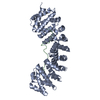
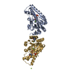
| Deposited unit | 
| ||||||||
|---|---|---|---|---|---|---|---|---|---|
| 1 |
| ||||||||
| Unit cell |
| ||||||||
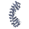
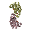
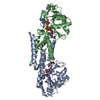
| Details | The biological assembly is a dimer in the asymmetric unit. |
-Components
| #1: Protein | Mass: 25086.713 Da / Num. of mol.: 2 / Mutation: L164F Source method: isolated from a genetically manipulated source Source: (gene. exp.) Homo sapiens (human) / Plasmid: pET15b / Production host:  Escherichia coli (E. coli) / References: UniProt: P78330, phosphoserine phosphatase Escherichia coli (E. coli) / References: UniProt: P78330, phosphoserine phosphatase#2: Chemical | 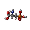  Type: D-peptide linking / Mass: 169.073 Da / Num. of mol.: 2 / Source method: obtained synthetically / Formula: C3H8NO5P Type: D-peptide linking / Mass: 169.073 Da / Num. of mol.: 2 / Source method: obtained synthetically / Formula: C3H8NO5P |
|---|
-Experimental details
-Experiment
| Experiment | Method: X-RAY DIFFRACTION / Number of used crystals: 1 |
|---|
- Sample preparation
| Crystal | Density Matthews: 2.47 Å3/Da / Density % sol: 50.14 % | ||||||||||||||||||||||||||||||||||||||||||
|---|---|---|---|---|---|---|---|---|---|---|---|---|---|---|---|---|---|---|---|---|---|---|---|---|---|---|---|---|---|---|---|---|---|---|---|---|---|---|---|---|---|---|---|
| Crystal grow | Temperature: 295 K / Method: vapor diffusion, hanging drop / pH: 7.2 Details: PEG 6000, pH 7.2, VAPOR DIFFUSION, HANGING DROP, temperature 295K | ||||||||||||||||||||||||||||||||||||||||||
| Crystal grow | *PLUS Temperature: 22 ℃ / pH: 5 | ||||||||||||||||||||||||||||||||||||||||||
| Components of the solutions | *PLUS
|
-Data collection
| Diffraction | Mean temperature: 100 K |
|---|---|
| Diffraction source | Source: SYNCHROTRON / Site: PAL/PLS  / Beamline: 6B / Wavelength: 0.9794 Å / Beamline: 6B / Wavelength: 0.9794 Å |
| Detector | Type: MACSCIENCE / Detector: IMAGE PLATE / Date: Jul 20, 2001 |
| Radiation | Monochromator: GRAPHITE / Protocol: MAD / Monochromatic (M) / Laue (L): M / Scattering type: x-ray |
| Radiation wavelength | Wavelength: 0.9794 Å / Relative weight: 1 |
| Reflection | Resolution: 2.51→30 Å / Num. all: 82983 / Num. obs: 15855 / % possible obs: 95.2 % / Observed criterion σ(F): 2 / Observed criterion σ(I): 2 / Biso Wilson estimate: 42.3 Å2 |
| Reflection shell | Resolution: 2.51→2.66 Å / % possible all: 75.4 |
| Reflection | *PLUS Highest resolution: 2.5 Å / Lowest resolution: 30 Å / Num. measured all: 82983 / Rmerge(I) obs: 0.034 |
- Processing
| Software |
| |||||||||||||||||||||||||
|---|---|---|---|---|---|---|---|---|---|---|---|---|---|---|---|---|---|---|---|---|---|---|---|---|---|---|
| Refinement | Method to determine structure: MAD / Resolution: 2.51→41.78 Å / Rfactor Rfree error: 0.011 / Data cutoff high absF: 1149683.08 / Isotropic thermal model: RESTRAINED / Cross valid method: THROUGHOUT / Stereochemistry target values: Engh & Huber
| |||||||||||||||||||||||||
| Solvent computation | Solvent model: FLAT MODEL / Bsol: 45.1554 Å2 / ksol: 0.314391 e/Å3 | |||||||||||||||||||||||||
| Displacement parameters | Biso mean: 33.6 Å2
| |||||||||||||||||||||||||
| Refine analyze |
| |||||||||||||||||||||||||
| Refinement step | Cycle: LAST / Resolution: 2.51→41.78 Å
| |||||||||||||||||||||||||
| Refine LS restraints |
| |||||||||||||||||||||||||
| LS refinement shell | Resolution: 2.5→2.66 Å / Rfactor Rfree error: 0.037 / Total num. of bins used: 6
| |||||||||||||||||||||||||
| Xplor file |
| |||||||||||||||||||||||||
| Refinement | *PLUS Highest resolution: 2.5 Å / Lowest resolution: 500 Å / % reflection Rfree: 7 % | |||||||||||||||||||||||||
| Solvent computation | *PLUS | |||||||||||||||||||||||||
| Displacement parameters | *PLUS | |||||||||||||||||||||||||
| Refine LS restraints | *PLUS
|
