 Movie
Movie Controller


+ Open data

- Basic information
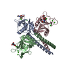
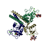
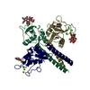
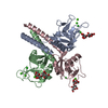
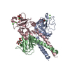
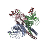
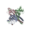
| Entry | Database: PDB / ID: 1kx0 | |||||||||
|---|---|---|---|---|---|---|---|---|---|---|
| Title | Rat mannose protein A (H189V I207V) complexed with man-a13-man | |||||||||
 Components Components | MANNOSE-BINDING PROTEIN A | |||||||||
 Keywords Keywords |  IMMUNE SYSTEM / SUGAR BINDING PROTEIN / LECTIN / C-TYPE LECTIN / CALCIUM-BINDING PROTEIN IMMUNE SYSTEM / SUGAR BINDING PROTEIN / LECTIN / C-TYPE LECTIN / CALCIUM-BINDING PROTEIN | |||||||||
| Function / homology |  Function and homology information Function and homology informationcalcium-dependent carbohydrate binding / complement activation, lectin pathway / oligosaccharide binding / killing by host of symbiont cells / collagen trimer / surfactant homeostasis / phosphatidylinositol-4-phosphate binding / polysaccharide binding / protein homotrimerization / D-mannose binding ...calcium-dependent carbohydrate binding / complement activation, lectin pathway / oligosaccharide binding / killing by host of symbiont cells / collagen trimer / surfactant homeostasis / phosphatidylinositol-4-phosphate binding / polysaccharide binding / protein homotrimerization / D-mannose binding / positive regulation of phagocytosis / multivesicular body / complement activation, classical pathway / calcium-dependent protein binding / protease binding / defense response to Gram-positive bacterium / calcium ion binding / protein homodimerization activity / extracellular space / identical protein bindingSimilarity search - Function | |||||||||
| Biological species |  Rattus norvegicus (Norway rat) Rattus norvegicus (Norway rat) | |||||||||
| Method | X-RAY DIFFRACTION / MOLECULAR REPLACEMENT / Resolution: 2 Å | |||||||||
 Authors Authors | Ng, K.K. / Kolatkar, A.R. / Park-Snyder, S. / Feinberg, H. / Clark, D.A. / Drickamer, K. / Weis, W.I. | |||||||||
 Citation Citation | Journal: J.Biol.Chem. / Year: 2002 Title: Orientation of bound ligands in mannose-binding proteins. Implications for multivalent ligand recognition. Authors: Ng, K.K. / Kolatkar, A.R. / Park-Snyder, S. / Feinberg, H. / Clark, D.A. / Drickamer, K. / Weis, W.I. | |||||||||
| History |
|
- Structure visualization
| Structure viewer | Molecule: MolmilJmol/JSmol |
|---|
- Downloads & links
-Download
| PDBx/mmCIF format | 1kx0.cif.gz | 110.8 KB | Display | PDBx/mmCIF format |
|---|---|---|---|---|
| PDB format | pdb1kx0.ent.gz | 88.8 KB | Display | PDB format |
| PDBx/mmJSON format | 1kx0.json.gz | Tree view | PDBx/mmJSON format | |
| Others |  Other downloads Other downloads |
-Validation report
| Arichive directory | https://data.pdbj.org/pub/pdb/validation_reports/kx/1kx0ftp://data.pdbj.org/pub/pdb/validation_reports/kx/1kx0 | HTTPS FTP |
|---|
-Related structure data
| Related structure data |  1kwtC  1kwuC  1kwvC 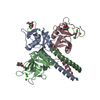 1kwwC  1kwxC 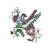 1kwyC  1kwzC  1kx1C  1kzaC  1kzbC 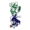 1kzcC 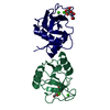 1kzdC  1kzeC C: citing same article ( |
|---|---|
| Similar structure data |


-Links
 PDBj
PDBj


- Assembly
| Deposited unit | 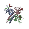
| ||||||||||
|---|---|---|---|---|---|---|---|---|---|---|---|
| 1 |
| ||||||||||
| Unit cell |
| ||||||||||
| Components on special symmetry positions |
|
-Components
| #1: Protein | Mass: 16425.631 Da / Num. of mol.: 3 / Fragment: residues 90-238 of P19999 / Mutation: H189V, I207V Source method: isolated from a genetically manipulated source Source: (gene. exp.) Rattus norvegicus (Norway rat) / Gene: MBL1 / Production host:  Escherichia coli (E. coli) / References: UniProt: P19999 Escherichia coli (E. coli) / References: UniProt: P19999#2: Polysaccharide | 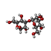  , Oligosaccharide / Class: Metabolism / Mass: 342.297 Da / Num. of mol.: 3 , Oligosaccharide / Class: Metabolism / Mass: 342.297 Da / Num. of mol.: 3Source method: isolated from a genetically manipulated source Details: oligosaccharide / References: 3alpha-alpha-mannobiose #3: Chemical | ChemComp-CA /   Mass: 40.078 Da / Num. of mol.: 9 / Source method: obtained synthetically / Formula: Ca Mass: 40.078 Da / Num. of mol.: 9 / Source method: obtained synthetically / Formula: Ca#4: Chemical | Chloride  Mass: 35.453 Da / Num. of mol.: 3 / Source method: obtained synthetically / Formula: Cl Mass: 35.453 Da / Num. of mol.: 3 / Source method: obtained synthetically / Formula: Cl#5: Water | ChemComp-HOH / | Water Mass: 18.015 Da / Num. of mol.: 370 / Source method: isolated from a natural source / Formula: H2O Mass: 18.015 Da / Num. of mol.: 370 / Source method: isolated from a natural source / Formula: H2O |
|---|
-Experimental details
-Experiment
| Experiment | Method: X-RAY DIFFRACTION / Number of used crystals: 1 |
|---|
- Sample preparation
| Crystal | Density Matthews: 3.3 Å3/Da / Density % sol: 62.77 % | ||||||||||||||||||||||||||||||||||||||||||||||||||||||||
|---|---|---|---|---|---|---|---|---|---|---|---|---|---|---|---|---|---|---|---|---|---|---|---|---|---|---|---|---|---|---|---|---|---|---|---|---|---|---|---|---|---|---|---|---|---|---|---|---|---|---|---|---|---|---|---|---|---|
| Crystal grow | Temperature: 298 K / Method: vapor diffusion, hanging drop / pH: 8 Details: 8-13% PEG 8000 or 3500, 100mM Tris-Cl pH=8.0, 10mM NaCl, 20mM Cacl2, 2mM NaN3. Protein solution: 12mg/ml in 10 mM NaCl, 10mM CaCl2, 200mM man-a13-man. VAPOR DIFFUSION, HANGING DROP at 298K | ||||||||||||||||||||||||||||||||||||||||||||||||||||||||
| Crystal grow | *PLUS Temperature: 20-22 ℃ | ||||||||||||||||||||||||||||||||||||||||||||||||||||||||
| Components of the solutions | *PLUS
|
-Data collection
| Diffraction | Mean temperature: 100 K |
|---|---|
| Diffraction source | Source: ROTATING ANODE / Type: RIGAKU / Wavelength: 1.5418 Å |
| Detector | Type: RIGAKU RAXIS IIC / Detector: IMAGE PLATE / Date: Jun 30, 1998 |
| Radiation | Monochromator: MIRRORS / Protocol: SINGLE WAVELENGTH / Monochromatic (M) / Laue (L): M / Scattering type: x-ray |
| Radiation wavelength | Wavelength: 1.5418 Å / Relative weight: 1 |
| Reflection | Resolution: 2→30 Å / Num. obs: 38640 / % possible obs: 93.2 % / Observed criterion σ(I): -3 / Rsym value: 0.046 |
| Reflection shell | Resolution: 2→2.05 Å / Rmerge(I) obs: 0.244 / % possible all: 92 |
| Reflection | *PLUS Lowest resolution: 30 Å / Redundancy: 2.5 % / Rmerge(I) obs: 0.046 |
| Reflection shell | *PLUS % possible obs: 92 % / Redundancy: 2.4 % |
- Processing
| Software |
| |||||||||||||||||||||||||
|---|---|---|---|---|---|---|---|---|---|---|---|---|---|---|---|---|---|---|---|---|---|---|---|---|---|---|
| Refinement | Method to determine structure: MOLECULAR REPLACEMENT / Resolution: 2→30 Å / Cross valid method: THROUGHOUT / σ(F): 0
| |||||||||||||||||||||||||
| Refinement step | Cycle: LAST / Resolution: 2→30 Å
| |||||||||||||||||||||||||
| Refine LS restraints |
| |||||||||||||||||||||||||
| Xplor file |
| |||||||||||||||||||||||||
| Refinement | *PLUS | |||||||||||||||||||||||||
| Solvent computation | *PLUS | |||||||||||||||||||||||||
| Displacement parameters | *PLUS | |||||||||||||||||||||||||
| Refine LS restraints | *PLUS
|
